 Sep 30 2026
 advanced imaging AI
Why Multimodal Prostate AI Needs More Than Image Labels: Annotation, Segmentation, and Clinical Data Quality
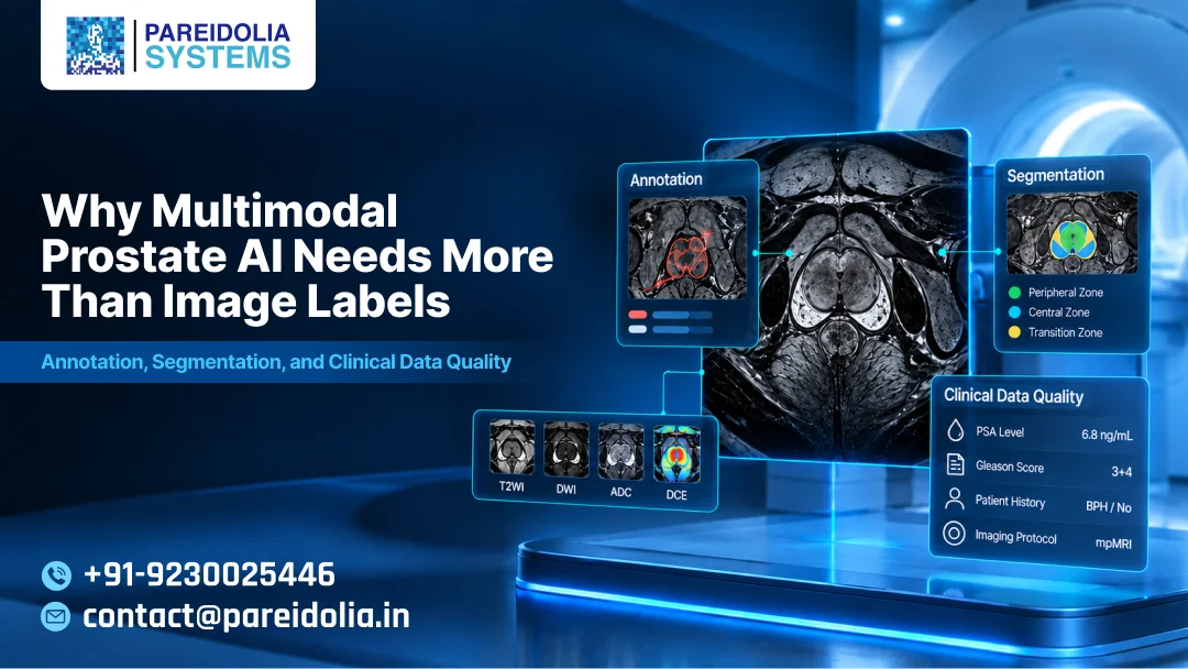
Prostate cancer diagnosis has always been a puzzle assembled from different pieces of evidence — a PSA number, an MRI sequence, a biopsy report, a pathologist’s Gleason score. No single image tells the whole story. That’s exactly why prostate AI has moved toward multimodal models: systems trained not on one scan type, but on the combination of T2-weighted MRI, diffusion-weighted imaging (DWI), apparent diffusion coefficient (ADC) maps, dynamic contrast-enhanced (DCE) sequences, and often clinical and pathology data layered on top.
The promise is real. A well-trained multimodal prostate AI model can flag clinically significant lesions earlier, reduce unnecessary biopsies, and support radiologists working through PI-RADS scoring under real-time pressure. But the promise only holds if the data feeding the model is built correctly. And this is where most multimodal prostate AI projects quietly run into trouble — not in the model architecture, but in the annotation pipeline behind it. It’s the layer of work Pareidolia Systems specializes in: turning raw, messy, multi-sequence imaging into AI-ready ground truth that healthcare teams can actually build on.
The Temptation to Treat It Like a Labeling Problem
It’s easy to think of AI development as a data-in, model-out process, where “getting the data ready” means drawing boxes or masks on images and moving on. For single-modality projects, that assumption can (mostly) survive. For multimodal prostate AI, it falls apart almost immediately.
Consider what a single prostate case actually involves: a T2 sequence showing anatomical detail, a DWI/ADC pair showing restricted diffusion, sometimes a DCE sequence showing enhancement patterns, and a radiology report scored against PI-RADS criteria. Each modality has a different resolution, a different slice thickness, and sometimes a different patient positioning between sequences. A lesion that is unambiguous on ADC can be nearly invisible on T2. Label each modality in isolation, without cross-referencing them, and you don’t get a multimodal dataset — you get several single-modality datasets that happen to share a patient ID.
This is precisely why Pareidolia’s workflow starts before any annotation happens at all. When a dataset comes in, the team’s first move isn’t to open an annotation tool — it’s to draft a Standard Operating Protocol (SOP) with the client, defining inclusion and exclusion criteria, zone definitions, and how discordant findings across sequences should be handled. That upfront structure is what keeps a multimodal dataset from becoming a set of disconnected single-modality labels.
Annotation: More Than Marking What’s Visible
Good annotation for prostate AI requires a defined protocol, not a well-intentioned annotator’s judgment call. Questions that need answering before a single pixel gets labeled include:
- Which zone does the lesion belong to — peripheral, transition, or central?
- Is the annotation being drawn per-slice or volumetrically across the full 3D stack?
- How should annotators handle lesions visible on DWI/ADC but ambiguous on T2, or vice versa?
- What’s the inclusion/exclusion criteria for equivocal PI-RADS 3 findings?
- How should the target scan be reconciled against the reference standard — biopsy location, whole-mount pathology, or follow-up imaging?
None of these questions have generic answers. They require annotators who understand prostate anatomy and PI-RADS scoring logic, not just annotators skilled with a segmentation tool. This is where Pareidolia’s model differs from generalist data-labeling vendors: its annotators are trained specifically for clinical imaging work and are regularly assessed for domain awareness across radiology sub-specialties — not treated as interchangeable labelers moved between unrelated projects. Clinical literacy on the annotation team is what actually determines dataset quality.
Segmentation: Where Precision Actually Gets Tested
Bounding boxes might be adequate for some detection tasks, but prostate AI models built for clinical use — especially anything supporting lesion volume estimation, treatment planning, or PSA density calculations — need proper segmentation: prostate gland contours, zonal boundaries, and lesion masks, ideally in 3D rather than slice-by-slice approximations. Medical image segmentation of this kind is one of Pareidolia’s core service lines, alongside annotation, 3D model creation, and dedicated quality control.
Segmentation quality has direct clinical consequences. An imprecise gland contour skews prostate volume, which skews PSA density, which is a metric urologists actually use in decision-making. A lesion mask that’s too generous inflates apparent lesion size; one that’s too conservative under-represents extracapsular extension risk.
This is also where inter-annotator agreement becomes essential rather than optional. Prostate lesions, especially at the PI-RADS 3 boundary, are genuinely ambiguous even to experienced radiologists. A dataset built from a single annotator’s interpretation inherits that annotator’s bias. Pareidolia treats inter-annotator agreement as a measured, tracked part of its process — adjudicating disagreements against a defined reference standard rather than leaving them to a single reviewer’s judgment — precisely because it’s one of the most overlooked steps in prostate AI pipelines built under deadline pressure.
Clinical Data Quality: The Layer Most Pipelines Forget
Image annotation alone still leaves gaps that only clinical data can fill. A multimodal prostate AI model trained purely on imaging, without structured clinical context, is working with one hand tied behind its back. PSA values and PSA density, prior biopsy history, & the actual histopathology outcome (Gleason grade group, tumor volume from whole-mount pathology) all sharpen what “ground truth” means for a given case.
Integrating this well requires consistent linkage between imaging studies, biopsy records, and pathology reports at the lesion level (not just the patient level), standardized terminology across sources that describe grading differently, and documented handling of discordant cases — for example, when imaging suggests a PI-RADS 4 lesion but targeted biopsy returns benign tissue. These are often the most clinically informative cases, & how a dataset handles them says a lot about its overall rigor.
This is where Pareidolia’s Quality Control service line does its work: structured review passes, spot-checks against the reference standard, and documented correction workflows that catch these issues before they reach a training set — not after a model’s performance plateaus unexpectedly. Being platform-agnostic is key. Our team works seamlessly within whatever annotation or PACS environment is available, eliminating the need for a costly or disruptive migration just to achieve consistent QC.
Why This Matters More in Prostate Imaging Than Almost Anywhere Else
Multimodal prostate AI sits in a particularly unforgiving spot. The disease is common enough that models need to generalize across large, diverse populations, but subtle enough in its imaging presentation that small annotation inconsistencies meaningfully affect performance. Add in that clinical decisions — biopsy or no biopsy, active surveillance or treatment — depend on the output, and the margin for error in the underlying data shrinks considerably.
What separates models that perform well in a paper from models that hold up in deployment is almost always the unglamorous groundwork — annotation protocols built around clinical reasoning, segmentation precision validated across annotators, and clinical data integrated with the same rigor as the imaging itself. This is the gap Pareidolia’s radiology-focused, cross-domain annotation expertise is built to close, backed by transparent, real-time updates through the annotation cycle so clients aren’t left waiting to find out where a project stands.
Building It Right From the Start
None of this means multimodal prostate AI needs to be slower or more expensive to get right. It means the annotation and data quality phase deserves the same deliberate planning as the model architecture does. A clear SOP defined up front, annotators with genuine familiarity with prostate imaging and PI-RADS criteria, measured inter-annotator agreement, and clinical data linked at the lesion level rather than bolted on afterward — these aren’t extras. They’re what determine whether a multimodal prostate AI model is a research curiosity or a tool clinicians can actually trust.
If your team is building or scaling a multimodal prostate AI pipeline & wants a partner who understands both the imaging and the clinical context behind it, Pareidolia Systems can help — from SOP drafting through annotation, segmentation, and quality control. Book a free demo to see how the workflow fits your dataset.