Ophthalmology

Ophthalmology

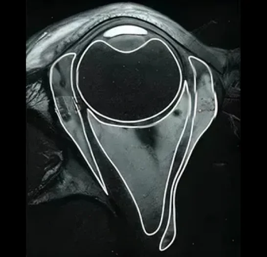
Pareidolia Systems LLP (the leading medical annotation company for deep learning and best medical image segmentation provider) specializes in expert outsourced medical image annotation for AI in Ophthalmology. We are dedicated to helping you create medical datasets for machine learning for critical ophthalmic imaging modalities, including CT, MRI, X-rays, ultrasound, and Dacryocystography. We generate high-fidelity AI training data for radiology startups and innovators by accurately labeling crucial anatomical structures—such as the globe, optic nerve, extraocular muscles, and orbital bones—along with detecting pathologies like foreign bodies, cystic lesions, and tumors.

Types of Medical Image Annotation in Ophthalmology

Ophthalmology X-ray Annotation & segmentation

Pareidolia Systems offers precise annotation of ophthalmic X-rays, primarily focusing on bony orbital structures and radiopaque foreign bodies. We annotate fractures, calcifications, and the presence and location of foreign objects within the orbit. While soft tissue visualization is limited on X-rays, our detailed annotations provide AI models with valuable datasets to assist in detecting relevant bony abnormalities and foreign body localization, aiding prompt clinical decision-making and surgical planning.

Dacryocystography Annotation

Pareidolia Systems provides specialized annotation for dacryocystography, an imaging technique used to visualize the lacrimal drainage system using contrast-enhanced X-rays or CT. We annotate key anatomical parts including the lacrimal sac, canaliculi, and nasolacrimal duct, as well as obstructions, stenoses, and filling defects caused by stones (dacryoliths) or inflammation. Our precise labeling supports AI models in diagnosing lacrimal drainage disorders and planning corrective surgeries.

CT Annotation & Segmentation for Ophthalmology

Our annotation workflow for ophthalmic CT scans includes detailed segmentation of orbital bones, globe, optic nerve, extraocular muscles, and adjacent sinuses. We identify fractures, foreign bodies, tumors, inflammations, and anatomical variants. With clinically validated annotations, AI models are enabled to deliver high-accuracy diagnoses and support surgical navigation for orbital and ocular conditions.

MRI Annotation & Segmentation for Ophthalmology

Pareidolia offers expert annotation of orbital and ocular MRI scans, emphasizing soft tissue contrast to delineate the optic nerve, ocular muscles, lacrimal gland, and orbital fat. We label pathologies such as optic neuritis, tumors, vascular malformations, and inflammatory processes.

Ophthalmic Ultrasound Annotation & Segmentation

Pareidolia Systems offers concise and precise annotation of ophthalmic ultrasound images, covering the globe, optic nerve, retinal layers, and orbital structures. We accurately delineate foreign bodies, intraocular masses, cysts, and vascular lesions. These expertly annotated datasets enable AI models to aid in diagnosing retinal abnormalities, tumors, vitreous hemorrhages, and more, supporting enhanced clinical decision-making with non-invasive, real-time imaging.

Contact us