 Jul 7 2026
 Advanced 3D Medical Image Segmentation
What Is the Difference Between image classification vs segmentation in Medical AI?
Why This Distinction Matters in Clinical AI?
When a radiologist reviews a chest CT or a pathologist examines a histology slide, they are doing two things simultaneously: recognising what is present and localising precisely where it is. Medical AI systems replicate this process through two distinct techniques — image classification vs segmentation.
Choosing the wrong technique for a clinical task does not just affect accuracy. It can mean the difference between a system that flags a disease and one that can actually guide a surgeon’s incision or measure tumour volume for treatment planning.
At Pareidolia Systems LLP, our work in healthcare AI is built on selecting the right analytical framework for each clinical context. This post breaks down image classification vs segmentation in plain terms — and explains why the choice is one of the most consequential decisions in medical AI development.
What Is Image Classification in Medical AI?
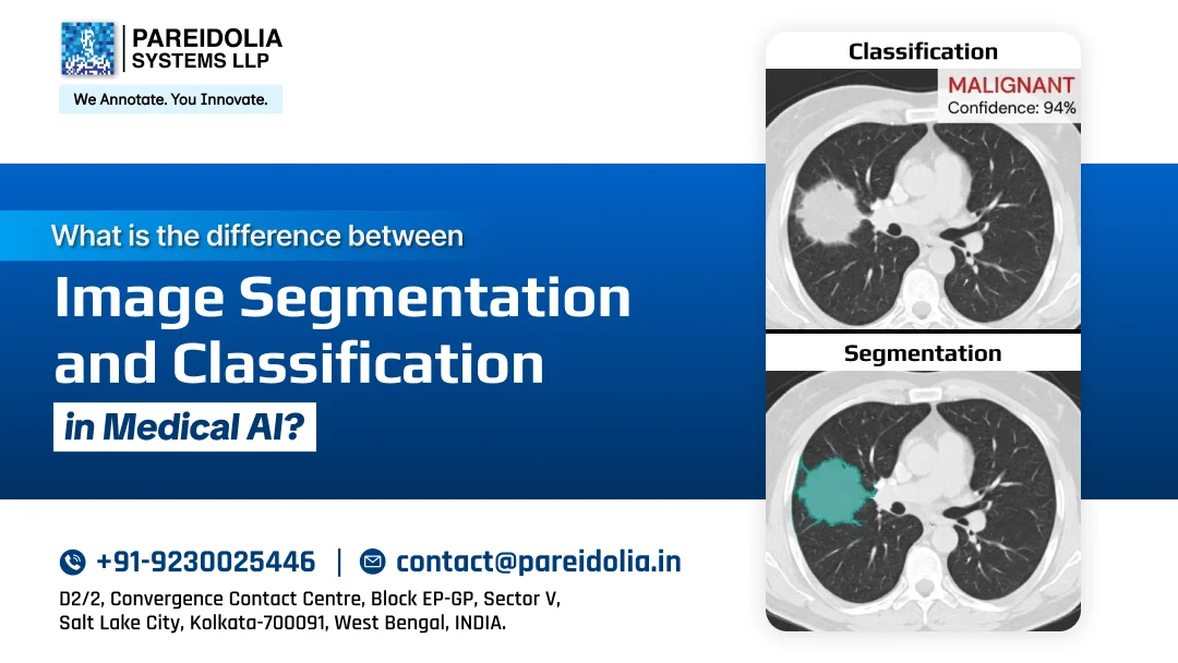
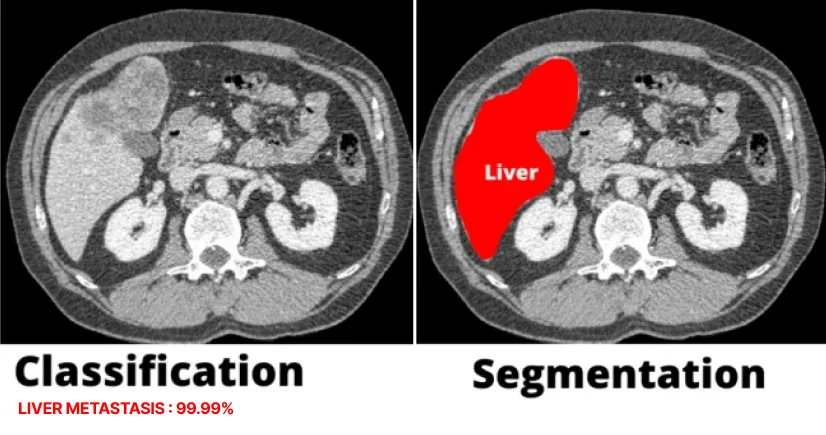
Image classification is the task of assigning a predefined label to an entire image or scan. The model takes the full image as input and outputs a category.
How It Works
A deep learning model (typically a Convolutional Neural Network or CNN) is trained on thousands of labelled images. It learns to extract features such as textures, shapes, intensity patterns, and maps those features to a class label.
Example outputs:
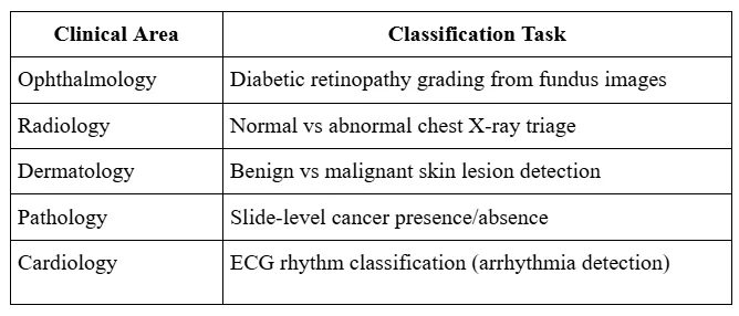
- “Diabetic retinopathy: Moderate”
- “Chest X-ray: Pneumonia positive”
- “Dermatoscopy: Melanoma suspected”
Where Classification Is Used in Medicine
Strengths of Classification
- Computationally lighter and faster to train
- Requires less granular annotation (image-level labels are sufficient)
- Ideal for screening and triage workflows where speed matters
- Well-suited to binary decisions (positive/negative, urgent/non-urgent)
Limitations of Classification
- Provides no spatial information — the model cannot tell you where the finding is
- Cannot measure lesion size, volume, or margin
- Offers limited interpretability — clinicians cannot see what the model is responding to without additional tools (e.g., Grad-CAM)
What Is Image Segmentation in Medical AI?
Image segmentation is the task of assigning a label to every pixel (or voxel, in 3D imaging) in a medical image. Instead of a single output label, the model produces a complete mask that outlines the region of interest.
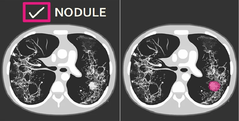
The Three Types of Segmentation Relevant to Medical AI
- Semantic Segmentation: Every pixel is assigned to a class (e.g., tumour tissue, healthy parenchyma, background). No distinction is made between individual instances of the same class. Use case: organ delineation for radiotherapy planning.
- Instance Segmentation: Like semantic segmentation, but individual objects of the same class are distinguished separately. Use case: counting and individually outlining polyps in a surgical video.
- Panoptic Segmentation combines semantic and instance segmentation into a single unified output. Use case: complex surgical scene understanding.
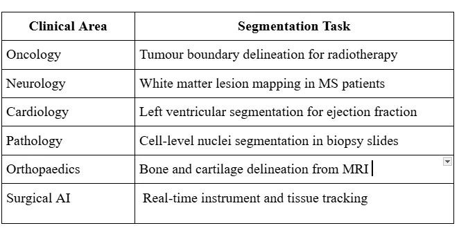
Where Segmentation Is Used in Medicine
Strengths of Segmentation
- Produces actionable spatial outputs — margins, volumes, boundaries
- Enables quantitative measurement (tumour volume, lesion load, organ size)
- Essential for treatment planning and surgical guidance
- Supports multimodal integration (e.g., aligning segmentation masks across CT, MRI, and PET)
Limitations of Segmentation
- Requires pixel-level annotations — far more time-intensive and expensive to create
- Computationally heavier (models like U-Net, Mask R-CNN, and nnU-Net are resource-intensive)
- More complex validation and quality assurance requirements
- Errors at boundaries can carry significant clinical consequences
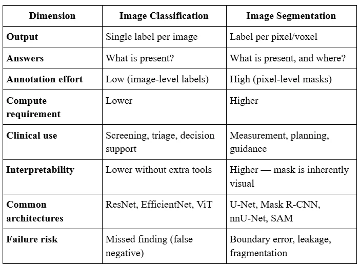
Image Classification vs Segmentation: Head-to-Head Comparison
The Middle Ground: Object Detection and Localisation
Between classification and segmentation sits object detection, which identifies where something is using a bounding box rather than a precise pixel mask. For many clinical tasks, counting nodules, flagging suspicious regions for radiologist review, offers a practical balance between annotation cost and spatial precision.
This is sometimes called weakly supervised localisation: a classification model is trained with image-level labels but is interpreted spatially using techniques like Class Activation Maps (CAM) or Grad-CAM to highlight the regions that drove the prediction.
How Pareidolia Systems Supports Classification and Segmentation Projects
At Pareidolia Systems LLP, we support medical AI development by delivering high-quality annotated datasets tailored to each client’s project requirements. Whether a project requires image classification, semantic or instance segmentation, 3D anatomical modelling, or a combination of these approaches, our workflows are designed to produce clinically reliable ground truth data for AI training and validation.
Our approach focuses on:
-
Project-specific annotation workflows
Classification requires accurate image-level labels, while segmentation requires precise pixel-level or voxel-level annotations. We customize annotation processes based on each client’s AI objectives.
-
Clinical accuracy and quality assurance
Our teams use domain expertise and structured quality checks to deliver reliable annotations across medical imaging applications, including radiology, pathology, and other specialties.
-
Advanced annotation capabilities
We support classification, semantic and instance segmentation, polygon annotations, landmark identification, volumetric segmentation, and 3D model creation to meet diverse medical imaging needs.
-
Data security and compliance
We follow stringent quality control and data protection practices aligned with HIPAA, GDPR, and applicable regulatory requirements to ensure secure handling of medical imaging data.
Pareidolia Systems LLP helps healthcare technology companies, researchers, and AI developers build reliable datasets for medical imaging AI applications through accurate annotation, quality control, and scalable workflows.
About Pareidolia Systems LLP
Pareidolia Systems provides clinically aware, multi-platform radiology annotation to help imaging AI teams offload edge-case validation and retrain models with flawless ground truth. We provide a cloud-native Enterprise Imaging Platform (VNA/PACS) and a unified ecosystem that offers a one-stop solution. We unify image acquisition across any modality with built-in de-identification and interoperable APIs to feed your applications seamlessly.
Explore our work: www.pareidolia.in
Contact us for a consultation: business@pareidolia.in