 May 13 2026
 3D Models in Medical Image Analysis
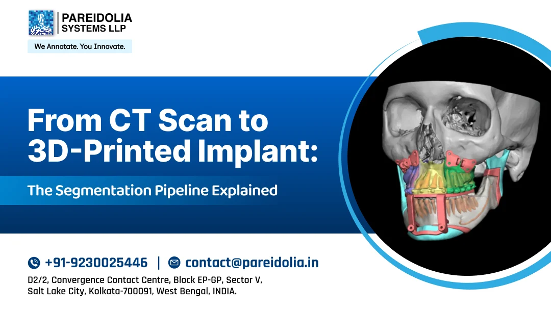
How 3D Printed Medical Implants Are Made: CT Scans to Implant Process
The healthcare sector is growing rapidly, particularly with the increased use of technologies such as 3D printing and advanced medical imaging, which are making diagnosis and treatment more accurate than ever before. The most viable innovation today is the 3D printed medical implants, which assist doctors in making implants that fit the exact anatomy of an individual patient.
But how does a simple CT scan turn into a perfectly fitted implant?
In simple terms, it all comes down to a step-by-step workflow involving 3D medical imaging, medical image segmentation, and advanced manufacturing. In this guide, we’ll walk you through the complete CT scan to implant process, along with practical insights from Pareidolia System LLP.
Understanding the Foundation: CT Scans and 3D Medical Imaging
It all starts with a CT scan, where X-rays are used to capture highly detailed cross-sectional images of the human body, which are then stacked together to form a precise digital structure.
This is where the use of 3D medical imaging becomes necessary. Doctors and engineers are able to view complex anatomical structures in three dimensions, rather than examining flat 2D slices. This is vital in the planning of the surgery, particularly in orthopaedic implants, craniofacial reconstruction, and cases of trauma, where precision is vital.
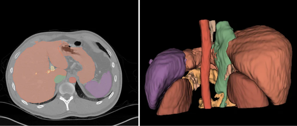
Raw CT data cannot, however, be directly used to create 3D printed medical implants. It contains multiple overlapping tissues such as bones, muscles, and organs. To extract only the required anatomical structure from the CT scan data for custom implant manufacturing, further processing is needed—this is where segmentation comes in.
What is Medical Image Segmentation?
Medical image segmentation is the procedure of isolating particular anatomical structures (such as bones, tissues, or tumours) from CT or MRI data. In simple terms, it works like digitally “cutting out” the exact region needed for building accurate 3D printed medical implants.
This step is one of the most critical parts of the CT scan to 3D model process, because even a small error in segmentation can lead to poor implant fit, affecting both functionality and surgical outcomes.
Segmentation can be performed using different approaches:
- Manual segmentation – Experts outline structures slice by slice for maximum control
- Semi-automatic methods – Software assists the process, but still requires human validation
- AI-based segmentation – Uses advanced algorithms to enhance speed and accuracy
As AI has rapidly grown in medical imaging, segmentation is now not only faster, scalable, and more accurate, but it is also significantly enhancing workflows in 3D printing in healthcare.
How 3D Printed Medical Implants Are Made
The creation of 3D printed medical implants follows a multi-step process:
- CT scan data acquisition
- Image preprocessing
- Medical image segmentation
- 3D reconstruction
- Implant design
- Validation and testing
- 3D printing
Every step ensures the accuracy and safety of the performance of the final implant.
Step 1: CT Scan Data Collection
It starts with high-resolution CT scans that have been stored in DICOM format and that are capable of capturing detailed anatomical structures.
Better scan quality directly improves the accuracy of the CT scan to the 3D model process and reduces errors in later stages.
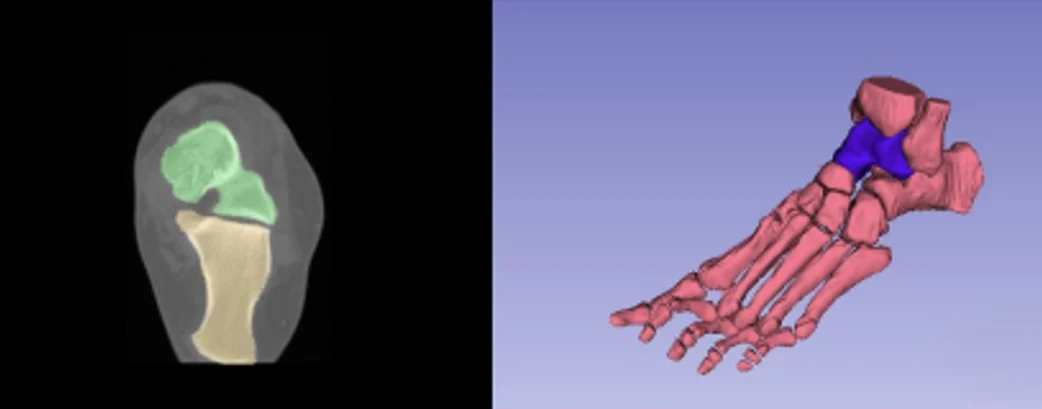
Step 2: Medical Image Segmentation
Medical image segmentation refers to the process of isolating a particular structure of the body, such as a bone, in the CT data.
This is also a very important step as it determines the exact shape of the 3D printed medical implants and makes sure that it fits the anatomy properly.
Step 3: From CT Scan to 3D Model Creation
The CT scan to 3D model process converts imaging data into a printable format.
Key Steps:
- Extract the segmented region
- Generate 3D mesh
- Convert to STL format
- Clean and optimise the model
The stage establishes the background for the correct 3D printed medical implants.
Step 4: Implant Design and Development
Using CAD tools, engineers design customised implants based on patient anatomy.
Important factors include fit, strength, and biocompatibility, ensuring that 3D printing in healthcare delivers safe and effective results.
Step 5: Validation and Testing
Before manufacturing, the implant undergoes validation, such as:
- Fit verification
- Structural analysis
- Surgical feasibility
This ensures the implant is ready for real-world clinical use.
Step 6: 3D Printing in Healthcare
The last design is then 3D printed in healthcare technologies.
Typical materials are titanium and polymer materials used in medicine, which means that complex and accurate structures of implants are possible.
How Pareidolia Systems powers the CT to Implant Workflow:
At Pareidolia Systems LLP, we follow a structured workflow to ensure accurate CT scan to 3D model conversion and implant-ready outputs.
We begin with high-quality DICOM data processing, followed by precise medical image segmentation to isolate anatomical regions. The data is then processed to result in optimized 3D models with appropriate mesh correction and validation.
We are interested in the accuracy, efficiency, and usability in the real world to ensure that the final models are reliable to be used in the 3D printing industry in the healthcare field.
Why Precision-Focused Teams Choose Pareidolia
- Moreover, pixel-perfect medical image segmentation is delivered with high attention to anatomical accuracy and structural detail for demanding healthcare AI and implant workflows.
- In addition, experienced and certified medical imaging specialists are trained in handling complex CT and MRI datasets across multiple anatomical regions and clinical applications.
- As a result, our expertise in CT/MRI processing, 3D reconstruction, and implant-ready model preparation supports patient-specific healthcare solutions.
- Additionally, clinically aligned segmentation workflows are designed to support precision-driven applications such as surgical planning, custom implant manufacturing, and medical AI development.
- Furthermore, rigorous quality control protocols focus on consistency, accuracy, and reliable output generation across every project.
- Moreover, scalable and responsive operational support, including 24×7 availability, enables faster turnaround times for global healthcare and medical technology teams.
- Overall, there is a strong focus on precision, consistency, and workflow adaptability to meet the requirements of complex medical imaging projects.
With hands-on experience in 3D medical imaging and implant workflows, the company focuses on delivering accurate and clinically relevant digital models for healthcare applications.
Common Mistakes in CT to Implant Workflow
Errors in the CT scan to implant process can significantly affect results.
Poor quality CT scans tend to result in an inaccurate model, whereas poor segmentation of medical images may result in distorted geometry of implants. Disregarding mesh defects can also lead to printing failures.
These are some of the mistakes that should be avoided in manufacturing quality 3D printed medical implants.
Best Software for Medical Image Segmentation
- Additionally, the accuracy of medical image segmentation and 3D medical imaging workflows is enhanced using the right tools.
- Popular software includes Materialise Mimics, 3D Slicer, and ITK-SNAP, which can support DICOM process and the generation of 3D models. These tools also offer more advanced features such as AI-assisted segmentation.
- The selection of the right software will guarantee effective and credible CT scan to 3D models processes.
Cost Breakdown of 3D Printed Medical Implants
The cost of 3D printed medical implants depends on several factors.
Major components include imaging, medical image segmentation, software processing, design, and manufacturing. Additionally, material selection also affects cost. Although there may be higher initial costs, 3D printing in healthcare tends to lower long-term surgical costs.
Real-World Applications
Applications of 3D printed medical implants include:
- Orthopedic implants
- Cranial reconstruction
- Maxillofacial and dental implants.
These applications enhance accuracy and patient outcomes in the current healthcare systems.
Benefits of 3D Printed Medical Implants
- Personalized treatment
- Faster recovery
- Improved surgical accuracy
- Better implant fit
- Reduced operation time
Future of 3D Printing in Healthcare
The future of 3D printing in healthcare includes:
- AI-driven automation
- Faster workflows
- Increased adoption in hospitals
With the advancement in technology, the demand for 3D printed medical implants will keep on growing.
Our Thoughts:
Overall, the process of manufacturing customised medical implants from raw CT scans is a strong illustration of how technology is revolutionising healthcare.
Furthermore, the process is increasingly becoming efficient and accessible due to the continuous innovation in AI in medical imaging and 3D medical imaging.
FAQ Section
1. What are 3D printed medical implants?
In simple terms, customised medical implants are 3D printed medical implants designed using a patient’s medical imaging data.
2. What is the application of the CT scans in the creation of implants?
Additionally, CT scans provide detailed internal images of the body, which are used to create accurate 3D models for implant design.
3. What is medical image segmentation?
In simple terms, medical image segmentation can be described as the act of isolating specific parts of the body from imaging data in order to accurately model them.
4. How is medical image segmentation done?
Firstly, the process of medical image segmentation is performed using dedicated software to isolate certain anatomical structures using the CT scan or MRI scan in order to properly model them using 3D modelling software.
5. What are 3D printed implants made out of?
Typically, materials such as titanium and biocompatible polymers are used because they are suitable for medical applications.
6. Are 3D printed implants safe?
Yes, they are safe and are in wide use in modern medical practice when designed and tested properly.
7. What is the importance of the quality of the CT scan to 3D printing?
As a result, good CT scans guarantee correct segmentation and accurate 3D models, which help minimise errors in implant design.
8. Why Partner with Pareidolia Systems LLP?
Because precision-driven healthcare AI and implant workflows demand more than standard annotation. They require pixel-perfect segmentation, experienced medical imaging specialists, and clinically reliable outputs at scale.