 Oct 14 2026
 Advanced 3D Medical Image Segmentation
Medical Image Annotation Services: Use Cases Across Healthcare AI
Ever wondered how a healthcare AI model spots a tiny lung nodule on a chest CT scan? Spoiler alert: it doesn’t “see” anything on its own.
AI only recognizes a potential tumor or lesion if it has analyzed thousands of previous scans, each one meticulously outlined, categorized, and verified by expert radiologists and medical annotators.
That is where medical image annotation service come in.
By converting raw DICOM images and X-rays into high-quality, structured training data, these specialized annotation teams bridge the gap between a flashy AI demo and a clinically dependable tool at the bedside.
In this guide, we’ll explore what medical image labeling actually involves, where it’s making the biggest impact across healthcare AI, and how to choose an annotation partner you can trust with sensitive clinical data.
What Is a Medical Image Annotation Service?
At its core, medical image annotation is the backbone of modern computer vision in healthcare. It’s the process of precisely labeling medical scans, including CT, MRI, X-ray, ultrasound, and mammography, so machine learning algorithms can accurately detect, measure, and classify complex anatomy and pathology.
In practice, expert annotators work alongside clinical specialists and radiology-literate reviewers to mark up complex data. Using specialized annotation platforms built specifically for medical formats like DICOM and NIfTI, they transform raw scans into structured, high-quality healthcare AI training data ready for model training, validation, and testing.
However, medical image labeling goes far beyond simply outlining a tumor. Building reliable medical AI requires:
- Clinical Consistency: Standardized labeling across thousands of multi-slice 3D volumes.
- Protocol Adherence: Strict alignment with established diagnostic guidelines.
- Data Security & Compliance: Enterprise-grade security to safeguard sensitive patient data at every step.
Medical Image Annotation vs. Segmentation: What’s the Difference?
If you’re building artificial intelligence or computer vision models for healthcare, you’ve likely come across medical image annotation and medical image segmentation. While these terms are frequently used interchangeably, they refer to two distinct steps in the data preparation pipeline.
In fact, most advanced medical AI applications rely on both to train accurate, clinical-grade algorithms. Here is a clear breakdown of how they differ and how to choose the right approach for your AI project.
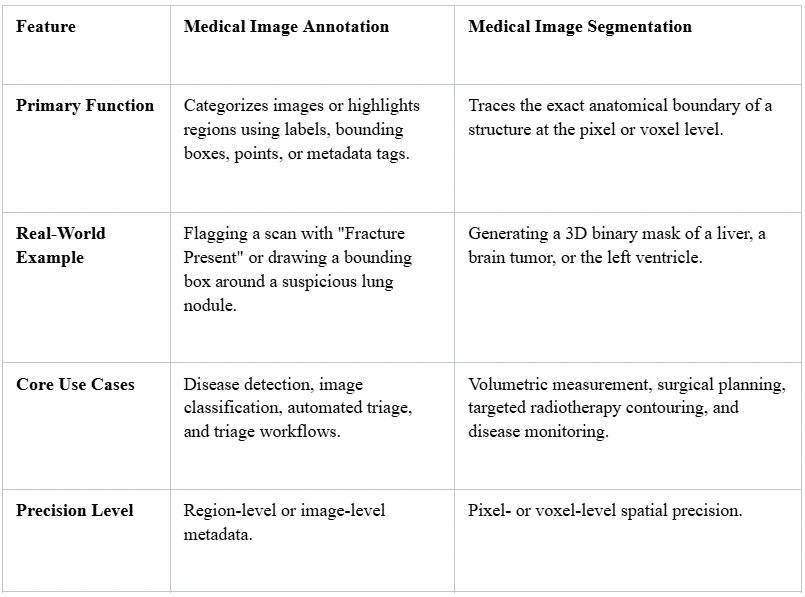
Medical Image Annotation vs. Segmentation
The Bottom Line for Healthcare AI Developers
To put it simply: Medical image labeling tells your AI model what is in a scan, while medical image segmentation tells it exactly where and how much is present.
If your machine learning model only needs to flag abnormalities or prioritize urgent scans for radiologist review, standard annotation is often fast, scalable, and sufficient. However, if your application needs to calculate precise measurements—such as tracking tumor volume growth, calculating cardiac ejection fraction, or measuring arterial plaque burden—pixel-accurate medical segmentation is mandatory.
Medical Image Annotation Across Imaging Modalities
Each modality has its own quirks, and a strong provider understands them.
- CT: High-resolution 3D volumes with hundreds of slices. Annotation needs efficient volumetric tools and attention to windowing.
- MRI: Multiple sequences (T1, T2, FLAIR, DWI) that show different tissue properties. Annotators often need to cross-reference sequences.
- X-ray: 2D projection images where overlapping structures make findings harder to isolate. Clear labeling guidelines are critical.
- Ultrasound: Operator-dependent, noisy and often in video or cine loops. Consistent labeling takes experienced reviewers.
- Mammography: Very subtle findings, such as microcalcifications and architectural distortion, in high-stakes screening settings. Quality control is non-negotiable.
Use Cases Across Healthcare AI
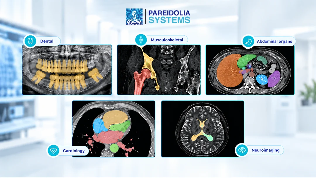
Here is how medical imaging annotation supports AI development across clinical specialties.
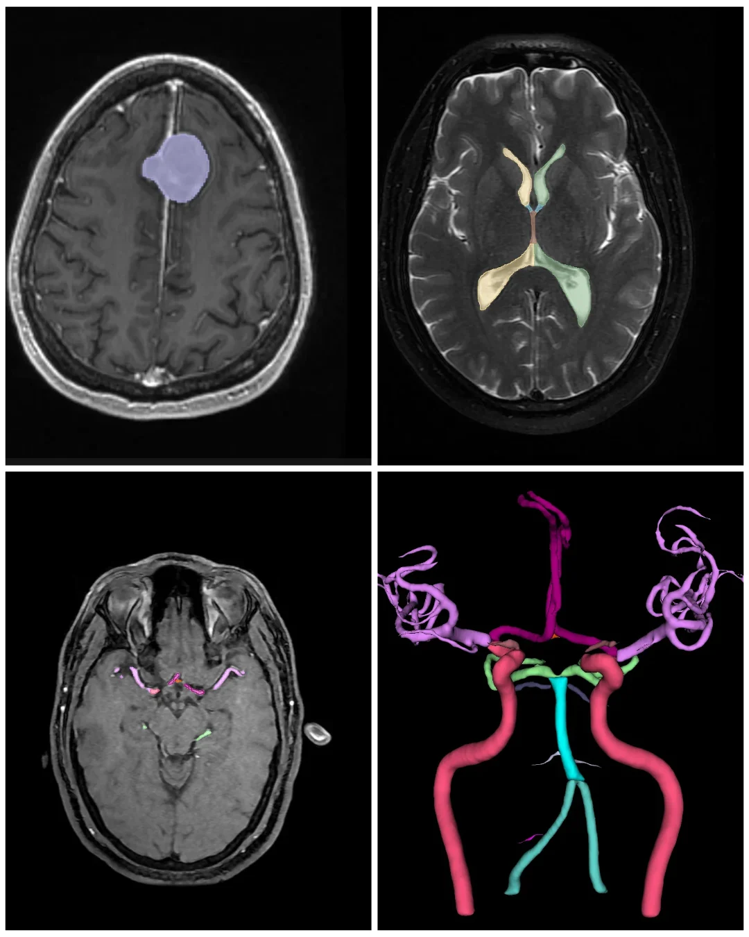
🧠 Neuroimaging
Brain MRI and CT are among the most data-hungry areas of healthcare AI. Annotation supports:
- Tumor and lesion segmentation (glioma, meningioma, metastases)
- Stroke and hemorrhage detection
- Multiple sclerosis lesion tracking
- Brain structure parcellation for neurodegenerative disease research
Neuroanatomy is subtle and varies widely between patients, so precision and consistency matter enormously.
❤️ Cardiovascular & Vascular
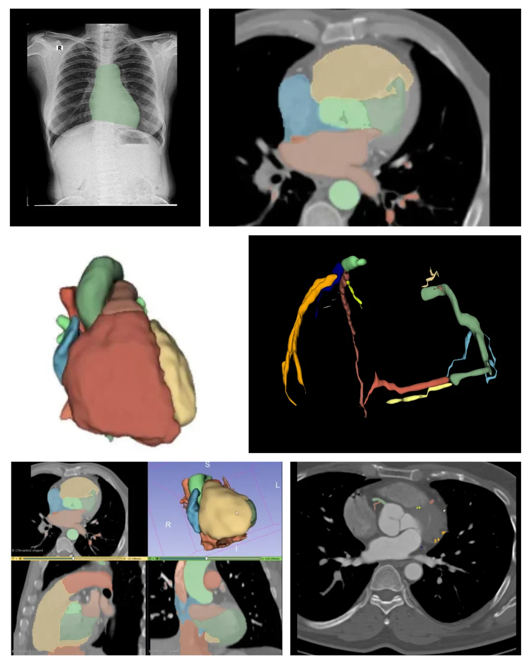
Heart and vessel imaging demands accuracy across time as well as space. Common annotation tasks include:
- Cardiac chamber and myocardium segmentation (CT, MRI, echo)
- Coronary artery and plaque labeling
- Aortic and vascular segmentation for aneurysm assessment
- Vessel centerline and stenosis marking
These datasets feed models that quantify cardiac function, flag disease early and support interventional planning.
🫁 Abdominal Organs
Abdominal CT and MRI contain many organs of varying shape and contrast. Annotation here covers:
- Liver, kidney, spleen, and pancreas segmentation
- Tumor and cyst delineation
- Organ volumetry for transplant and surgical planning
- Multi-organ labeling for general-purpose foundation models
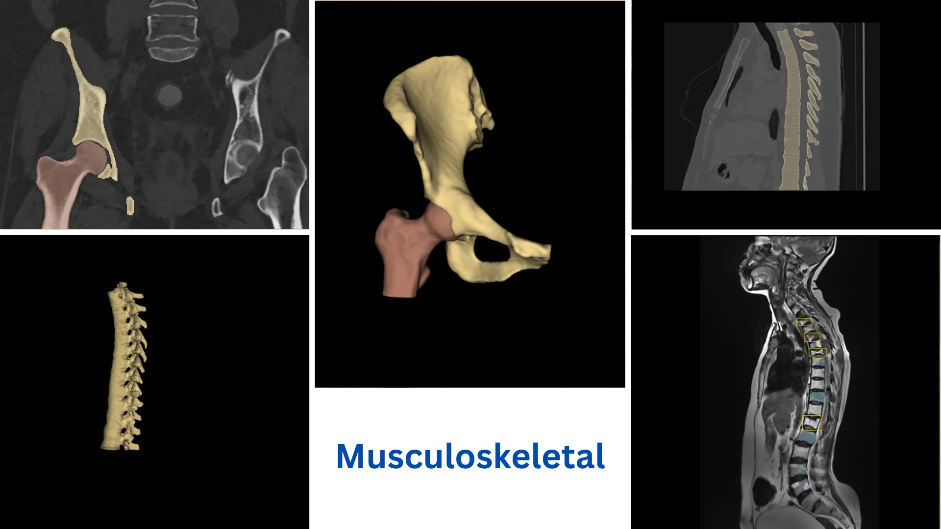
🦴 Musculoskeletal
From trauma to chronic joint disease, MSK imaging benefits greatly from precise labels:
- Fracture detection and localization
- Bone, cartilage and muscle segmentation
- Spine labeling (vertebrae, discs, canal)
- Joint assessment for orthopedic and sports medicine applications
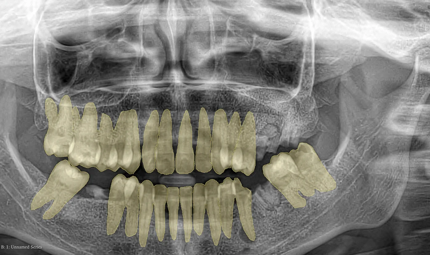
🦷 Dental
Dental AI is one of the fastest-growing applications of computer vision in healthcare. Typical annotations include:
- Tooth numbering and instance segmentation
- Caries, periapical lesion, and bone loss detection
- Implant planning and nerve canal marking
- Panoramic, bitewing and CBCT labeling
Why Annotation Quality Makes or Breaks Healthcare AI
A model is only as good as its labels. In healthcare AI data annotation, that isn’t just a best-practice guideline, it is the ultimate rule for building production-ready medical technology.
When annotation quality suffers, the model suffers. Poor medical data labeling creates severe downstream issues:
- Inconsistent segmentation boundaries: Models end up training on statistical noise rather than actual anatomical structures.
- Missed radiological or clinical findings: Unlabeled anomalies translate directly into dangerous false negatives in deployment.
- Severe label bias: Inconsistent inputs produce AI models that fail when exposed to different imaging scanners, patient populations, or hospital networks.
- Costly project delays: Stalled training runs often force teams to completely discard and re-label weak, low-quality datasets.
In real-world clinical deployment, the consequences extend far beyond dropping a few points on a benchmark score. High-precision medical datasets yield trustworthy diagnostic algorithms for clinicians and clear, audit-ready documentation for regulatory approval (like FDA 510(k) clearances).
Achieving high-quality medical annotation at scale requires a systematic pipeline: clear consensus guidelines, medically trained annotators, multi-stage QA reviews, inter-annotator agreement (IAA) metrics, and credentialed expert sign-offs on edge cases.
What Makes Pareidolia Different?
At Pareidolia, we treat medical data annotation as a clinical-grade discipline—not a commodity task. We bridge the gap between complex raw imaging and production-ready AI models.
- Purpose-Built for Healthcare: Custom workflows engineered specifically for CT, MRI, X-ray, ultrasound, and mammography never retrofitted from generic labeling tools.
- Deep 2D & 3D Segmentation: High-precision volumetric mapping across neuro, cardiovascular, abdominal, musculoskeletal, and dental subspecialties.
- Clinician-Led Expertise: Board-certified radiologists and medical specialists driving high inter-annotator agreement through standardized SOPs.
- Layered Quality Assurance: Multi-tiered QC protocols ensuring sub-pixel accuracy and clinical validity for every dataset.
- Native Medical Format Support: Seamless handling of DICOM, NIfTI, and structured annotation exports without data corruption or loss.
- Platform-Agnostic Integration: Complete ecosystem flexibility fluent in Redbrick AI, Mimics, 3D Slicer, CVAT, and custom client stacks.
- Enterprise Security & Compliance: HIPAA-compliant, HIPAA-aligned de-identification, and strict data governance protocols built into every pipeline.
- Scalable Turnaround: Predictable delivery timelines designed to scale seamlessly from proof-of-concept pilots to massive enterprise datasets.
How to Choose a Medical Image Annotation Provider
Selecting the right annotation vendor directly impacts your AI model’s accuracy, safety, and time-to-market. Before signing a contract, evaluate providers using this 8-step checklist:
1. Verified Modality Expertise: Do they have proven experience with your specific imaging modality (CT, MRI, X-ray) and anatomical target? Request sample datasets first.
2. Clinician-Led Labeling: Are certified radiologists or medical experts annotating and reviewing the data, or are they relying on generic crowdsourced workers?
3. Rigorous QA & Agreement Metrics: How do they quantify precision? Look for multi-tier quality assurance, consensus scoring, and active error-correction loops.
4. Native 3D Volumetric Capability: Can they handle complex multi-sequence and volumetric segmentation (e.g., DICOM/NIfTI), or are they limited to basic 2D bounding boxes?
5. Strict Security & HIPAA Compliance: What protocols protect patient privacy? Require end-to-end data encryption, robust access controls, and strict de-identification processes.
6. SOP & Protocol Adaptability: Will they strictly adapt to your custom labeling guidelines and help refine them as edge cases arise?
7. Proven Scalability & Pilot Testing: Can they maintain inter-annotator agreement as dataset volumes grow? Always run a paid pilot project before committing at scale.
8. Pipeline-Ready Tooling & Formats: Do their outputs integrate seamlessly into your AI pipeline, supporting platforms like Redbrick AI, CVAT, or 3D Slicer without custom reformatting?
FAQ: Medical Image Annotation Service
What is a medical image annotation service?
Medical image annotation service involve labeling and segmenting clinical scans (CT, MRI, X-ray, ultrasound) to generate ground-truth training data for healthcare AI and machine learning models.
What is the difference between medical image annotation and segmentation?
Annotation identifies and tags features, while 2D/3D segmentation outlines exact pixel or voxel boundaries of anatomical structures and lesions .
Why is precise annotation critical for healthcare AI?
Algorithm accuracy depends on label quality. High-precision clinical annotations directly improve model sensitivity, specificity, and generalization across diverse patient populations.
Which imaging modalities can be annotated?
Services cover all major modalities, including CT, MRI, X-ray, ultrasound, mammography, and specialized dental imaging.
Who performs medical image annotation?
Annotation is performed by specialized medical annotators and validated by board-certified radiologists or clinical experts using dedicated DICOM-native tools.
What are the main use cases for medical image segmentation?
Key applications include tumor delineation, organ volumetry, cardiac chamber analysis, vascular mapping, radiotherapy planning, and orthopedic structural labeling.
How is medical annotation quality ensured?
Top providers enforce strict SOPs, multi-tiered QC reviews, inter-annotator agreement metrics, radiologist audits, and proof-of-concept (PoC) pilot phases.
How is patient data protected during the annotation process?
Reputable partners utilize fully de-identified DICOM files, end-to-end encrypted pipelines, strict access controls, and adherence to HIPAA and GDPR standards.
Ready to Build Better Healthcare AI?
Your algorithm is only as strong as its training data. Avoid costly rework and accelerate clinical validation with expert-led data annotation.
Talk to the Pareidolia team. Partner with Pareidolia to scale your medical AI pipeline. Contact our team today to request a custom PoC or launch a pilot project.