 Mar 26 2025
 Scientific
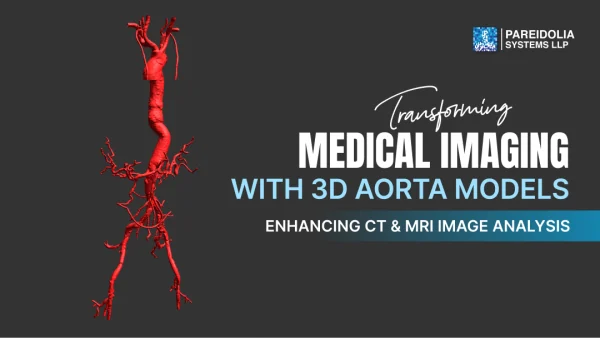
Transforming Medical Imaging with 3D Aorta Models: Enhancing CT & MRI Image Analysis
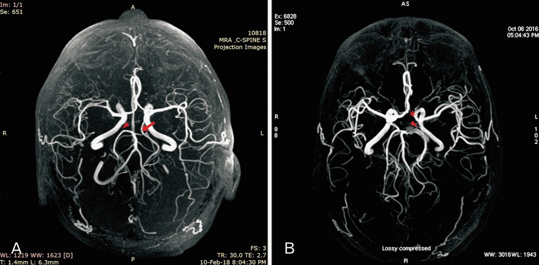
Medical imaging has evolved significantly, and one of the most groundbreaking advancements is the creation of 3D models from CT and MRI scans. Their 3D aorta models enable near-perfect accuracy and valuable insights, making them indispensable in the diagnosis and study of intricate vascular structures. The aorta, the body’s largest blood vessel, is critical for circulatory health. Turning a 2D image into a 3d image does just that, creating a high-detail 3D representation of a human body, allowing human/AI to gain more knowledge on the anatomical and pathological structure, accurately recognizing functions of cells and tissues in a place, thus allowing optimized treatment and answering in evolutionary dynamic pathways.
In this article, we will discuss state-of-the-art 3D aorta modeling that has changed the course of aorta diagnosis and research and how our advanced segmentation and 3D modeling services for CT and MRI images are bringing such an accuracy that has never been seen before in medical professionals. Our experienced team offers economical, accurate, and highly scalable solutions, assisting healthcare leaders in training AI-ML models for the end goal of improving diagnostics and patient outcomes.
Importance of 3D Aorta Models in Medical Imaging
The aorta plays an essential role in the circulatory system, transporting oxygenated blood from your heart to the rest of your body. Aneurysms, stenosis, or dissections all of which have potentially dire consequences. CT scans and MRIs offer critical information but interpreting these images can be difficult given the complexity of the aortic anatomy.
Benefits of 3D Aorta Models:
- Enhanced Visualization: 3D models offer a comprehensive view of the aorta, allowing precise assessment of its shape, size, and abnormalities.
- Accurate Measurements: Precise aortic diameter, length, and volume measurements assist in diagnosing conditions like aneurysms and stenosis.
- Pre-Surgical Planning: Surgeons can use 3D models to visualize abnormalities and plan procedures with greater accuracy, minimizing risks.
- Longitudinal Monitoring: 3D models facilitate accurate tracking of disease progression for chronic conditions affecting the aorta.
Transforming 2D imaging into detailed 3D representations unlocks vital diagnostic data, improving accuracy and treatment effectiveness.
The Power of 3D Modeling in CT & MRI Imaging
Creating a 3D aorta model from CT and MRI scans involves precise segmentation and reconstruction processes.
1. Segmentation:
Segmentation isolates the aorta from surrounding tissues using sophisticated algorithms that define its boundaries. Our team employs advanced techniques such as thresholding and region-growing algorithms to ensure accuracy in CT and MRI scans.
2. 3D Reconstruction:
After segmentation, specialized software transforms the data into a realistic 3D model, offering multiple viewing angles. This enhances pre-surgical planning, patient education, and AI-driven research.
Our cutting-edge software ensures high-resolution 3D models that are both accurate and adaptable for various applications in the healthcare industry.
How 3D Models Support AI & ML in Healthcare
The integration of 3D modeling with artificial intelligence (AI) and machine learning (ML) is transforming healthcare. AI-ML algorithms benefit significantly from high-precision 3D models, enabling faster, more reliable diagnostics.
Key AI-ML Applications:
- Automated Disease Detection: AI models trained with 3D data can identify aortic aneurysms, stenosis, and dissections with greater accuracy.
- Predictive Analytics: Machine learning algorithms use 3D models to assess disease progression, improving treatment decision-making.
- Surgical Guidance: AI-powered models recommend optimal surgical approaches based on high-accuracy 3D aorta reconstructions.
Through our services, healthcare innovators have developed AI-ML models capable of automating aortic disease assessment, leading to faster diagnoses and improved treatment plans.
Role of 3D Aorta Models in Medical Imaging
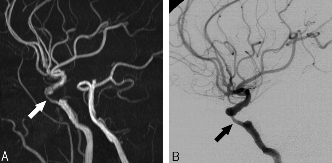
As the main highway for blood flow, the aorta carries oxygen-rich blood away from the heart and around the body. Even detecting abnormal findings such as aneurysms, stenosis, & dissections early in the way is critical to avoiding life-threatening scenarios. Having been inspired by routine imaging modalities such as CT and MRI which deliver practitioners with considerable information (as digital images, nevertheless remaining 2D), we acknowledge the limitations of this type of data when it comes to visualization and analysis.
Benefits of 3D Aorta Models:
Improved Visualization: An intact, high-resolution 3D model enables medical professionals to view the aorta from different perspectives, enhancing diagnostic accuracy.
Precise Measurements: The aorta’s diameter, volume, and shape can all be accurately calculated with 3D models, thus allowing for effective planning of treatment.
Pre-Surgical Planning Surgeons can simulate procedures to reduce risks and enhance patient outcomes.
Tracking the Course of Disease: Diseases of the aorta are often chronic, and their progression can be tracked over time more accurately.
The Process: 3D Models from CT & MRI Images Segmentation:
The first step is segmentation, where specialized algorithms delineate the aorta from surrounding tissues, using images from CT or MRI scans. For accurate 3D representation, high precision is crucial, and we only achieved this using advanced techniques such as thresholding and region-growing algorithms.
3D Reconstruction:
After segmentation, extracted data is used to produce the detailed 3D model using advanced medical imaging software. The model is applicable for surgical planning, AI training, and research purposes.
The 3D Aorta Models and Incorporation of AI & Machine Learning
Combining AI and machine learning with 3D aorta models is transforming the healthcare sector. When training AI models on realistic 3D representations, the medical imaging technology is enhanced in some of the following ways:
Privacy and Data Protection: AI in Healthcare AI can anonymize patient data to maintain privacy.
Predictive Analytics: Machine learning algorithms analyze 3D models and predict potential disease progression.
Surgical Assistance: Overall AI tools help in improving guidance during surgical operations that help decrease the risk of injury.
Why Choose Our 3D Aorta Modeling Services?
- Expertise & Precision: Our experienced team specializes in medical image analysis, segmentation, and 3D reconstruction, delivering highly accurate results.
- Cost-Effective Solutions: We offer affordable, scalable services to help organizations access advanced 3D models within budget constraints.
- High Accuracy: Precision is critical in medical imaging. Our meticulous segmentation and 3D modeling processes ensure true-to-life anatomical representation.
- Fast Turnaround: We provide timely 3D model delivery, supporting urgent healthcare research and clinical projects.
Real-World Impact: Collaborating with Leading Healthcare Companies
We have successfully partnered with top healthcare and research organizations, delivering high-quality 3D aorta models derived from CT and MRI scans. Our collaborations have advanced AI-ML research, enabling automated detection and analysis of aortic diseases, improving diagnostics, and refining surgical planning.
The Future of Healthcare: 3D Aorta Models & AI Integration
The ability to generate accurate 3D models from CT and MRI scans represents a significant breakthrough in cardiovascular diagnostics and treatment planning. By providing advanced segmentation, precise 3D reconstructions, and AI-ML training support, our services enable healthcare professionals to achieve earlier disease detection, better treatment planning, and improved patient outcomes.
If you’re seeking cost-effective, accurate, and scalable 3D aorta modeling services to enhance your healthcare research or AI development, we are here to help. Contact us today to learn more about how our expert solutions can support your medical imaging and AI initiatives.