 May 15 2025
 3D printing in neurosurgery
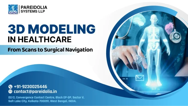
3D Modeling in Healthcare: From Scans to Surgical Navigation
Introduction: Why Imaging Data Is a Game-Changer
3D Modeling in Healthcare is revolutionizing diagnostics and treatment planning by enhancing visualization and precision across clinical workflows. The global medical imaging market was valued at approximately USD 41.6 billion in 2023 and is projected to grow at a CAGR of about 5–6% over the next decade. Advances like AI-powered segmentation, 3D model creation, VR & AR-guided Surgical navigation are driving this expansion—transforming scan data into surgical intelligence.
What Pareidolia Systems LLP Offers
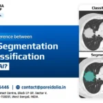
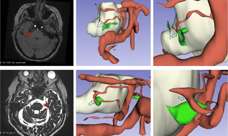
1. Medical Image Segmentation & Annotation
Using advanced Software platforms, we precisely segment CT/MRI scans—annotation & segmentation organs, vessels, and tissues down to the pixel. This pick-by-pixel accuracy ensures that your 3D models are anatomically reliable and clinically actionable, meaning every organ, tissue, and vessel is represented with high fidelity. Such precision allows for improved diagnostics, surgical planning, and AI model training, enabling clinicians and researchers to make informed, confident decisions based on accurate anatomical structures.
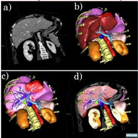
2. 3D Model Creation & Printing
We convert segmented imaging into virtual 3D models and optionally produce patient-specific anatomical 3D models. These digital replicas aid surgeons in visualizing and rehearsing complex procedures, improving strategy, reducing surprise, and enhancing outcomes.
3. AR-Guided Surgical Navigation
We integrate high-resolution 3D anatomical models into augmented reality platforms enabling surgeons to visualize patient-specific anatomy in real time during procedures. This holographic overlay is spatially registered to the patient’s body, Allowing for intuitive, heads-up navigation without relying solely on traditional imaging monitors. By aligning virtual anatomy with physical landmarks, AR significantly enhances intraoperative accuracy. Particularly in complex procedures such as biopsies, ablations, and orthopedic surgeries. Surgeons can perform needle placements and instrument guidance with greater confidence and spatial awareness.
4. VR-Guided Surgical Navigation
We convert segmented 3D anatomical data into immersive VR environments, allowing surgeons to explore patient-specific anatomy before entering the operating room. Using VR headsets, clinicians can rehearse procedures, assess spatial relationships, and plan optimal surgical paths. This approach improves depth perception, enhances anatomical understanding, and supports better decision-making—especially in complex cases like neurosurgery or cardiac interventions. VR-guided planning has been shown to reduce surgical risks, improve precision, & shorten operation times.
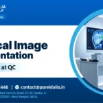
5. Quality Control in Medical Imaging
Each image, model, segmentation, and annotation is subjected to a multi-stage quality control process, including automated checks and manual reviews by trained experts. We maintain over 98% annotation accuracy and consistency rates, verified against gold-standard datasets. All outputs are validated in alignment with DICOM compliance protocols and institutional imaging governance. Ensuring every deliverable meets stringent clinical and regulatory benchmarks.
Key Benefits of Working with Us
Enhanced Surgical Precision
By overlaying real-time anatomical data, AR delivers sub-millimeter accuracy for procedures involving the skull base, spine, and abdomen.
Time & Cost Efficiency
Fewer instrument passes and less radiation exposure translate into faster operations, fewer complications, and shorter hospital stays.
Improved Clinical Outcomes
Surgeons report better anatomical insights and operative confidence—leading to fewer errors and improved patient recoveries.
Better Training & Collaboration
Using VR and collaborative 3D spaces, surgeons and trainees can practice procedures and enhance learning through realistic, hands-on simulations.
Compliance & Consistency
We maintain strict adherence to segmentation and imaging standards, ensuring uniformity across multi-site projects.
Why the Future Is 3D
3D Modeling in Healthcare is reshaping the future of surgery, diagnosis, and medical training. Here’s why it’s rapidly becoming the new standard in modern healthcare:
- Enhanced Visualization: 3D models offer a more accurate and comprehensive view of complex anatomical structures, allowing for better surgical planning and precision.
- Improved Surgical Accuracy: When integrated with AR/VR, 3D guidance enables sub-millimeter accuracy in instrument navigation, reducing the risk of damage to critical tissues.
- Reduced Complications: Real-time 3D navigation helps minimize tissue trauma, leading to fewer complications and faster patient recovery.
- Personalized Treatment: Patient-specific 3D models support tailored surgical approaches, improving outcomes across diverse organ systems.
- Better Training & Collaboration: VR-based 3D environments create immersive learning experiences for students and allow surgical teams to rehearse complex procedures collaboratively.
- Data-Driven Decisions: 3D insights improve preoperative planning by combining imaging data with real anatomical structures for informed clinical decisions.
3D Modeling in Healthcare is not just innovation—it’s transformation for safer, smarter, and more personalized care.
FAQs
Q1: What modalities do you support?
A: X-ray, CT, MRI, PET, and ultrasound imaging are all supported for annotation, segmentation, and 3D reconstruction.
Q2: How accurate are the segmentations?
A: Our pixel-level annotation and segmentation are validated against gold standards and clinician review. Our pixel-level annotation and segmentation validated against gold standards & clinician reviews. Making them ideal for diagnostic and surgical applications.
Q3: Do you offer physical 3D prints?
A: No. We only offer medical image segmentation and annotation services. We understand that precision and speed are crucial when transforming medical imaging data into accurate 3D-printed models. Our team of certified medical professionals is available 365 days a year, 24/7, ensuring you receive expert support whenever you need it—no delays, no compromises.
Q4: Can you support multi-center clinical trials?
A: Yes. All segmentation, annotation, and 3D models follow standardized protocols, ensuring quality and regulatory compliance across sites.
From pixel-precise segmentation, annotation, and 3D Modeling in Healthcare to immersive AR & VR surgical navigation. Pareidolia Systems LLP delivers end-to-end annotation and segmentation services for 3D healthcare models—engineered for precision, safety, and clinical efficacy. Partner with us to transform simple scan data into structured surgical intelligence.