Sep 8 2025
 3D Models in Medical Image Analysis
The Role of 3D Models in Medical Image Analysis: Improving Patient Outcomes and Surgical Precision
In today’s rapidly advancing healthcare industry, 3D medical imaging plays a vital role in diagnosing diseases, planning treatments, and guiding surgeries. Traditionally, medical imaging relied on two-dimensional (2D) scans, which often limited a clinician’s ability to fully visualize the complex anatomy of the human body. However, with the advent of 3D modeling in medical image analysis, healthcare professionals now have access to highly detailed, interactive representations of patient anatomy.
This innovation is revolutionizing the way doctors interpret imaging data, leading to better diagnoses, more precise surgeries, and significantly improved patient outcomes. Moreover, it is reshaping how medical image processing is applied in clinical trials, where accuracy and consistency are critical. Whether it’s medical imaging analysis or creating precise 3D models through advanced annotation, Pareidolia Systems LLP empowers healthcare innovators with accurate data-driven solutions.
What is Medical Imaging and Why is It Crucial?
Medical imaging is the process of creating visual representations of the body’s internal structures using advanced technologies such as:
-
- Magnetic Resonance Imaging
- Computed Tomography
- Positron Emission Tomography
- X-rays
- Ultrasound
These imaging techniques allow physicians to diagnose pathologies, monitor disease progression, and plan medical procedures. However, while 2D imaging has been standard for decades, it presents limitations when interpreting complex anatomical structures. For instance, surgeons must mentally reconstruct a 3D view from multiple 2D slices, which can lead to inaccuracies.
The integration of 3D medical imaging eliminates this challenge by providing lifelike models that can be rotated, sliced, and viewed from different angles—offering a far superior understanding of patient anatomy.
Evolution from 2D to 3D: A Game-Changer in Healthcare
For many years, doctors relied solely on 2D scans to assess injuries, plan surgeries, and detect abnormalities. While effective, 2D imaging lacked depth perception, which is crucial for understanding intricate anatomical relationships.
With 3D modeling, healthcare professionals now benefit from:
- Greater visualization: Viewing structures from every angle for better diagnosis.
- Improved accuracy: Identifying exact tumor sizes, bone fractures, or vascular abnormalities.
- Enhanced pre-surgical planning: Practicing procedures virtually before operating on patients.
This shift from 2D to 3D has been particularly impactful in specialties like neurosurgery, orthopedics, cardiology, and oncology, where precision is paramount.
How 3D Models Improve Patient Outcomes
1. Enhanced Diagnostic Accuracy
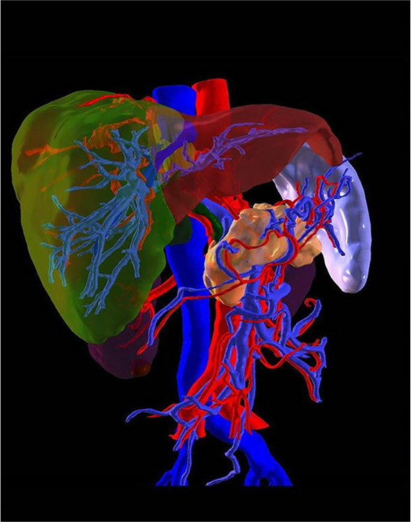
3D imaging allows doctors to detect abnormalities earlier and measure them with extreme precision. For instance, when diagnosing tumors, 3D visualization helps identify not only the size and location but also the proximity to critical blood vessels and nerves, minimizing risks during surgery.
2. Personalized Treatment Planning
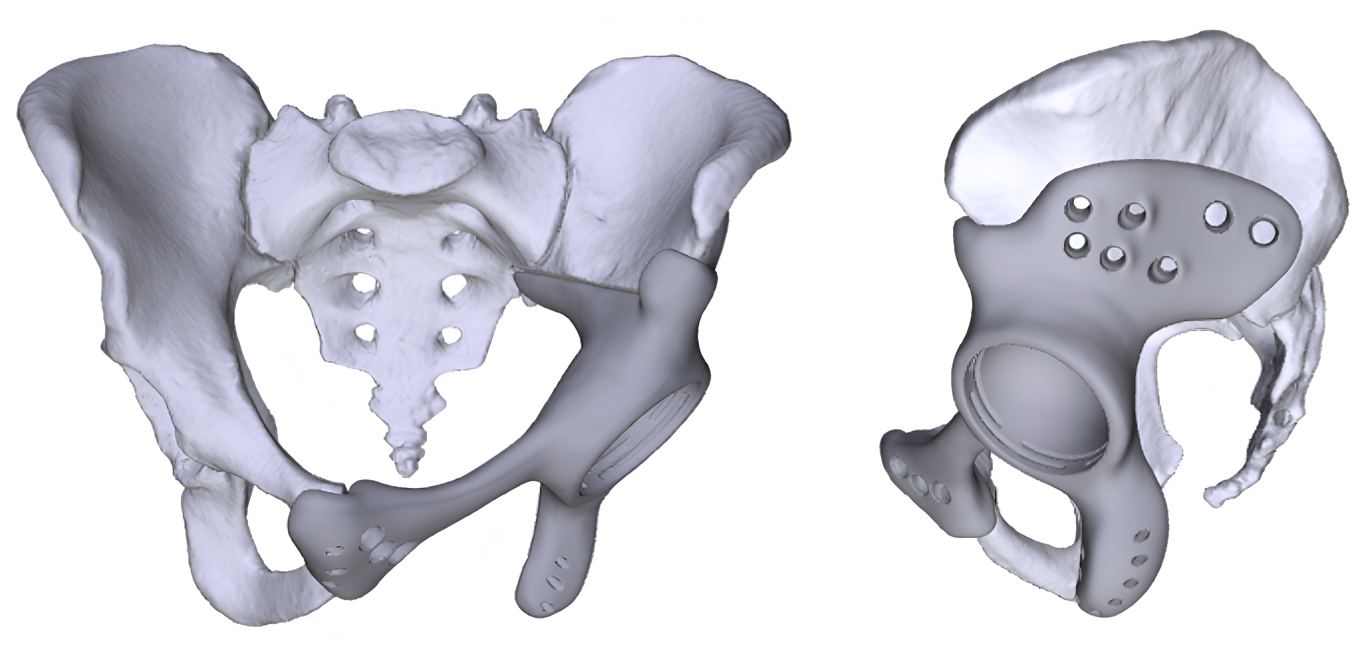
Every patient is unique, and 3D modeling supports personalized medicine by enabling treatment plans tailored to specific anatomical structures. Whether it’s fitting a customized implant or determining the safest surgical approach, 3D imaging significantly improves treatment effectiveness.
3. Reduced Surgical Risks
Complex surgeries often carry high risks due to limited visibility of internal structures. With 3D models, surgeons can simulate procedures beforehand, anticipate complications, and practice precise movements, reducing errors and recovery time.
4. Better Patient Communication
3D visualizations simplify explanations, helping patients understand their diagnosis, treatment plan, and surgical procedures clearly—leading to better compliance and confidence.
The Role of Medical Image Annotation in 3D Modeling
High-quality medical image annotation is the cornerstone of accurate 3D modeling in modern healthcare. At the intersection of innovation and compliance, expert annotation transforms raw medical scans into precise, structured data fueling breakthroughs in 3D printing, clinical research, and AI-driven diagnostics.
- 3D printing companies depend on detailed, anatomically correct annotations to create customized surgical models and patient-specific implants.
- Clinical research organizations (CROs) require consistently labeled data to ensure trial accuracy, regulatory alignment, and faster go-to-market timelines.
- AI healthtech companies leverage expertly annotated datasets to train robust, regulation-ready algorithms for diagnostics and treatment planning.
Our annotation services are designed for speed, accuracy, and full compliance with HIPAA, GCP, and GDPR, ensuring data integrity, patient safety, and scalable results.
3D Imaging in Clinical Trials
Clinical trials demand high levels of precision to evaluate the effectiveness of new treatments. Imaging in clinical trials has evolved significantly with the introduction of 3D modeling. Key benefits include:
- Accurate Measurement: Volumetric analysis of tumors or lesions provides precise data on treatment effects.
- Consistency Across Sites: Standardized imaging protocols ensure reliable data across multiple trial locations.
- Faster Approval Processes: Enhanced accuracy reduces trial errors, helping drugs and devices reach the market sooner.
Pareidolia Systems LLP: Driving Innovation in 3D Medical Image Analysis
At Pareidolia Systems LLP, we recognize that the future of healthcare lies in the intelligent use of data. Medical imaging produces vast amounts of information, but without precise interpretation and structured processing, its true potential often remains underutilized. This is where Pareidolia plays a pivotal role.
- Accurate Data Annotation & Segmentation
We transform raw medical imaging into structured datasets by carefully annotating and segmenting scans, ensuring accuracy for AI and clinical use. - 3D Modeling for Better Visualization
Our advanced 3D models help doctors and surgeons clearly visualize complex anatomical structures, aiding in early diagnosis and precise surgical planning. - AI-Driven Insights
By integrating machine learning, we enable faster, more consistent diagnostics while reducing human error, directly improving patient outcomes. - End-to-End Healthcare Solutions
From raw imaging data to AI-ready 3D models, we provide complete solutions that empower healthcare providers and accelerate medical technology innovation.
How 3D Models Enhance Surgical Precision
Surgeons across various specialties benefit immensely from 3D imaging technology. Some key applications include:
- Neurosurgery
3D models map the brain with incredible accuracy, helping neurosurgeons navigate delicate regions while avoiding critical nerves and blood vessels.
- Orthopedics
Custom implants designed with 3D imaging fit patients perfectly, reducing recovery time and improving mobility after surgery.
- Oncology
Cancer treatment planning relies on precise tumor mapping, allowing targeted radiation therapy while sparing healthy tissues.
- Cardiology
3D imaging enables visualization of congenital heart defects, aiding minimally invasive surgeries and improving patient outcomes.
Future Trends in 3D Medical Imaging
The future of medical imaging is being shaped by advancements in Artificial Intelligence (AI), Augmented Reality (AR), and Virtual Reality (VR). Emerging trends include:
- Real-Time 3D Imaging: Allowing surgeons to view dynamic anatomical changes during procedures.
- Predictive Modeling: Using AI to forecast disease progression and surgical outcomes.
- Immersive Surgical Simulations: VR-enabled 3D models help surgeons practice complex operations before entering the operating room.
The integration of 3D models in medical imaging is transforming healthcare by improving diagnostic accuracy, enhancing surgical precision, and advancing clinical trials. At Pareidolia Systems LLP, we provide end-to-end solutions in data annotation, segmentation, and 3D modeling, ensuring raw imaging data is converted into accurate, high-quality datasets for informed clinical decisions. Our advanced processing helps doctors visualize complex anatomical structures clearly, enabling early detection, effective treatment planning, and better patient communication. Beyond diagnostics, our solutions support AI-driven research and clinical trials, where annotated imaging data is vital for developing next-generation technologies. By combining expertise in AI innovation and healthcare software development, Pareidolia turns complex imaging into reliable insights, empowering providers to deliver precision-driven care and improved patient outcomes.