 Oct 16 2025
 AI in Breast Cancer Imaging
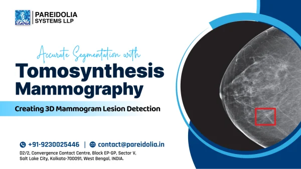
Accurate Segmentation with Tomosynthesis Mammography:Creating 3D Mammogram Lesion Detection AI
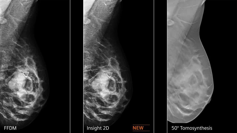
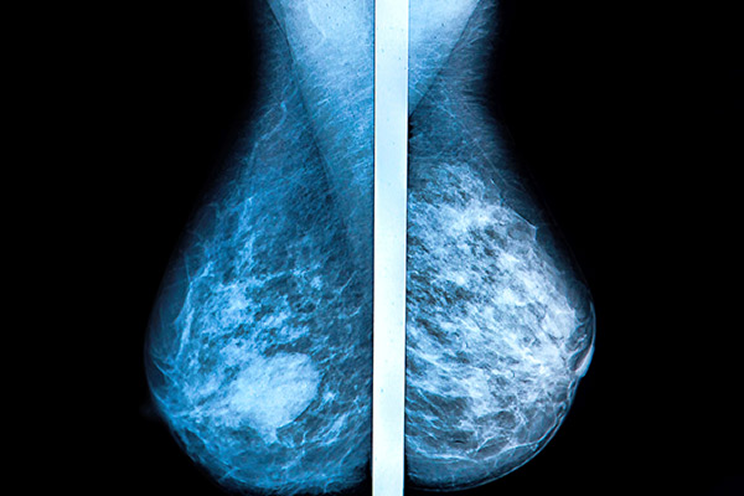
Breast cancer remains one of the most prevalent and life-threatening diseases affecting women globally. Early detection is crucial for improving survival rates, and technological advancements in medical imaging have transformed the diagnostic landscape. Among these innovations, digital breast tomosynthesis (DBT), commonly known as 3D mammography, has become a cornerstone for the precise detection of breast lesions.
The vast volume and complexity of 3D mammogram data present new challenges for radiologists. Manual interpretation is time-consuming and subjective, often leading to variability in results. To address this, Pareidolia Systems LLP focuses on precise segmentation of tomosynthesis mammography, which is essential for building robust AI-based 3D mammogram lesion detection. Through data annotation, segmentation, and 3D modeling, Pareidolia enables the development of AI systems that support radiologists in identifying lesions with greater accuracy, consistency, and speed.
Understanding Tomosynthesis Mammography
Digital breast tomosynthesis (DBT) is an advanced form of mammography that captures multiple low-dose X-ray images of the breast from different angles. These slices are reconstructed into a 3D image, providing a clearer view of internal structures compared to traditional 2D mammograms.
This 3D imaging approach minimizes the issue of tissue overlap, which often obscures small tumors or lesions in 2D scans. DBT significantly improves cancer detection rates, particularly in women with dense breast tissue.
Each tomosynthesis scan produces hundreds of image slices, making manual analysis labor-intensive. This is where AI-based lesion segmentation and detection offer revolutionary assistance.
What Is Tomosynthesis Mammography Segmentation?
Tomosynthesis mammography segmentation refers to the process of identifying and delineating regions of interest, such as lesions, masses, or calcifications, from 3D breast imaging data.
Segmentation converts raw pixel image information into structured, labeled data that AI systems can understand and analyze. It enables:
- Accurate localization of potential abnormalities.
- Quantitative measurement of lesion size, shape.
- Classification of density.
- Enhanced visualization for radiologists and researchers.
- Empowering to train on AI-based diagnostic models.
At Pareidolia, we ensure that every segmentation of tomosynthesis mammography images is the foundation upon which reliable AI models for 3D mammogram lesion detection can be built.
Pareidolia’s Expertise in 3D Mammogram Segmentation & Annotation
Pareidolia specializes in medical image annotation and segmentation for healthcare applications. Our approach combines technical precision, clinical understanding, and ethical data handling to ensure accuracy at every level of the imaging workflow.
1. Data Curation and Preparation
To ensure consistency prior to segmentation, imaging datasets are carefully curated by systematically removing all inconsistent data. This process requires a thorough quality check across all parameters, with special emphasis on identifying images vital for artifact removal and assessing the impact of the exposure factor.
2. Accurate Lesion Annotation
Our medical annotation experts meticulously label breast lesions, microcalcifications, and architectural distortions within DBT slices. Each annotation is guided by radiological protocols to ensure clinical validity and consistency.
3. 3D Segmentation and Reconstruction
Pareidolia performs pixel-level segmentation across multiple image slices and reconstructs the lesion in 3D volumetric form. This provides both visual and quantitative data crucial for developing AI models for breast lesion classification.
4. Quality Assurance
Every dataset undergoes multi-stage quality checks, combining automated verification with expert validation. Our emphasis on accuracy ensures that AI models trained with our data can achieve reliable performance in real clinical scenarios.
How AI Transforms 3D Mammogram Lesion Detection
AI integration in pathology detection in tomosynthesis mammography has revolutionized breast cancer screening and diagnosis. By learning from Pareidolia’s precisely annotated datasets, AI systems can assist in multiple clinical tasks:
1. Automated Lesion Detection
AI algorithms trained on precisely segmented tomosynthesis data can automatically identify and mark suspicious regions, allowing radiologists to focus on critical cases faster. This leads to earlier detection and shorter diagnostic times.
2. Enhanced Diagnostic Confidence
AI-driven systems reduce observer variability and provide quantitative lesion metrics, such as size, boundary irregularity, and contrast features, which support more objective diagnoses.
3. Improved Workflow Efficiency
With hundreds of images per scan, manual interpretation can be time-consuming. Automated 3D mammogram lesion detection AI streamlines the workflow, helping radiologists process more cases without compromising accuracy.
4. Data-Driven Treatment Planning
Accurate lesion segmentation provides valuable insights for treatment planning, such as determining surgical margins or monitoring tumor response during therapy. This data also supports longitudinal studies and clinical trials.
Deep Learning for Mammogram Lesion Detection
Deep learning plays a pivotal role in AI-based 3D mammogram analysis. Convolutional neural networks (CNNs) and transformer-based architectures can identify subtle features in breast images that may not be visible to the human eye.
Pareidolia’s segmented datasets provide the foundation for training such deep learning models. These datasets include:
-
Precisely segmented lesion boundaries.
-
Differentiation between benign and malignant structures.
-
Consistent labeling across imaging modalities and patient cases.
Through this approach, Pareidolia helps research organizations and medical institutions accelerate the development of AI for breast cancer imaging — ensuring data integrity and clinical relevance.
Clinical Impact of Tomosynthesis Mammography Segmentation
The clinical impact of tomosynthesis mammography segmentation extends far beyond automation. It directly enhances patient outcomes by making diagnostics more reliable, data-driven, and accessible.
1. Early and Accurate Detection
Precise segmentation allows AI to detect subtle lesions that might otherwise go unnoticed. Early detection leads to better treatment success rates and reduced patient morbidity.
2. Reduced False Positives and Negatives
By clearly distinguishing between normal tissue and potential abnormalities, AI-based 3D mammogram analysis reduces diagnostic errors and unnecessary biopsies.
3. Personalized Patient Care
Detailed lesion segmentation provides radiologists with insights into lesion morphology and progression, enabling personalized treatment planning and follow-up strategies.
4. Research and Clinical Innovation
High-quality segmentation data supports AI model development, validation, and multi-institutional collaboration.
Pareidolia’s End-to-End Process: From Image to AI Model
Pareidolia Systems LLP follows a structured multi-stage process that ensures the integrity of every AI development cycle.
-
- Stage 1: Data Acquisition and Curation
Sourcing de-identified DBT datasets that meet ethical and clinical standards.
- Stage 1: Data Acquisition and Curation
-
- Stage 2: Annotation and Labeling
Applying expert lesion markings and image categorization based on medical imaging protocols.
- Stage 2: Annotation and Labeling
-
- Stage 2A: Segmentation and 3D Modeling
Performing 3D breast imaging segmentation and volumetric reconstruction to visualize lesion boundaries in three dimensions.
- Stage 2A: Segmentation and 3D Modeling
-
- Stage 3: Enable Intelligent Breast Imaging AI: Through meticulous annotation and volumetric segmentation of breast lesions, we provide datasets that empower AI models to achieve reliable, accurate, and clinically meaningful detection in digital breast tomosynthesis.
-
- Stage 4: Clinical Implementation and Trials
Collaborating with hospitals and diagnostic centers for real-world validation of AI in breast cancer imaging.
- Stage 4: Clinical Implementation and Trials
Each stage demonstrates Pareidolia’s commitment to merging precision data with real clinical value.
Challenges in 3D Mammogram Segmentation and Pareidolia’s Solutions
1. High Data Volume
Tomosynthesis produces hundreds of slices per breast, making annotation challenging. Pareidolia addresses this high expert team with expert clinical review, improving both efficiency and accuracy.
2. Variability in Breast Density
Dense tissue can obscure lesions. Pareidolia ensures consistent results by training annotators on density-specific segmentation protocols and using AI-assisted image enhancement techniques.
3. Lesion Diversity
Lesions differ widely in texture, size, and morphology. Our adaptive segmentation workflows account for these variations, ensuring reliable results across diverse patient populations.
4. Maintaining Data Privacy
All datasets undergo strict anonymization and encryption to meet international privacy standards. Pareidolia ensures full compliance with healthcare data protection laws.
Why Precision Matters: Pareidolia’s Perspective
Precision segmentation goes beyond technical achievement — it is the bridge between imaging and insight. At Pareidolia Systems LLP, every dataset is a step toward transforming medical imaging into actionable clinical intelligence. Pareidolia empowers AI developers, AI Companies, Clinical Innovation Organizations, and healthcare institutions to detect, diagnose, and treat breast cancer with unprecedented precision.
Tomosynthesis mammography segmentation represents the future of breast cancer imaging — where AI, data annotation, and 3D modeling converge to improve diagnostic precision.
In the mission to fight breast cancer, every pixel matters — and at Pareidolia, we make every pixel count.
Our philosophy is rooted in accuracy, reliability, and collaboration: “We Annotate. You Innovate.”