 Nov 6 2026
 medical image analysis
Scaling Neurological AI Development Through Multimodality Brain Image Segmentation: CT, MRI, DSA & Beyond
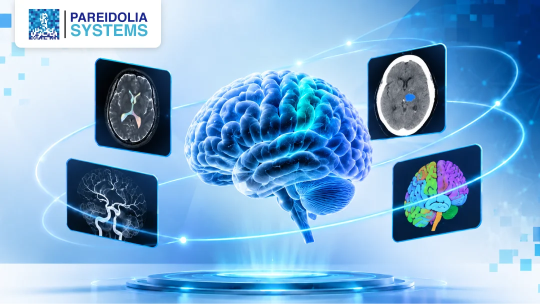
Neurological AI is only as good as the data it learns from, and for brain imaging that means precise, consistent, clinically meaningful segmentation across every modality a clinician relies on. Multimodality brain image segmentation is the process of outlining brain structures and pathologies across different imaging types, such as CT, MRI, DSA, PET, SPECT and fluoroscopy, so machine learning models can learn from a complete clinical picture.
The Growing Demand for Neurological Imaging Datasets
Healthcare AI developers and SaMD teams are building tools to detect brain tumours, triage stroke, flag aneurysms and track neurodegenerative change. Each of these tools needs large, accurately labelled datasets and the quality of those labels directly shapes how well a model performs once it reaches clinical use.
The challenge is that brain imaging is among one of the hardest areas to annotate well. Anatomy is intricate, pathology is subtle and no single modality tells the whole story. A CT scan may confirm a bleed within minutes, an MRI may reveal the soft-tissue detail around it, and a DSA may show exactly how blood is flowing through the affected vessels. Teams that train on only one view often leave clinical value on the table.
This is why more AI developers are turning to specialist partners for multimodality segmentation. In this blog, we explain why it matters, how each modality contributes, where it applies in neurological AI, and how Pareidolia Systems delivers AI-ready brain imaging datasets.
Why Multimodality Segmentation Matters?
Single-modality datasets can teach a model to recognise a pattern, but they rarely teach it to understand a patient. Three challenges explain why multimodality work needs a more careful approach.
Anatomical complexity: The brain packs grey and white matter, ventricles, deep nuclei, cortical regions and an intricate vascular network into a small space. Boundaries are often faint, and structures differ in appearance from one modality to the next. Segmenting them correctly requires real anatomical knowledge, not just tool skills.
Image variability: Scanner vendors, acquisition protocols, slice thickness, contrast phases and patient motion all change how a study looks. Add differences between hospitals and populations, and the same lesion can appear very different from one dataset to another. Models trained on narrow, inconsistent data tend to struggle when they meet new images.
Annotation consistency: When several annotators work on one project, small differences in how they interpret a boundary can multiply across thousands of studies. Without a clear SOP, defined inclusion and exclusion criteria, and structured review, label noise creeps in and quietly limits model performance.
Multimodality segmentation addresses these problems by combining complementary information, such as structure from CT and MRI, vessel dynamics from DSA and metabolic or perfusion data from PET and SPECT, under one consistent annotation framework.
Our Multimodality Segmentation Capabilities
Pareidolia Systems supports the full range of imaging types used in neurological AI development. Each modality brings its own strengths, and our annotators are trained to handle its specific challenges.
CT Brain Segmentation
CT scan is the first-line modality in emergency neurology because it is fast and widely available. We segment haemorrhages, infarcts, ventricles, skull and bone structures, midline shift and other findings used in stroke triage and trauma models. At Pareidolia, our experienced medical imaging team tackles complex anatomichallenges & edge cases through precise annotation, segmentation and expert-led quality validation.
MRI Brain Segmentation
MRI offers the soft-tissue detail needed for tumours, white matter lesions and neurodegenerative research. We work across sequences such as T1, T2, FLAIR, DWI, SWI, and contrast-enhanced series, segmenting tumour sub-regions, oedema, lesions and anatomical structures while keeping labels aligned between sequences.
DSA Segmentation
Digital subtraction angiography is the reference view for cerebral vasculature. We annotate vessels, aneurysms, stenoses, malformations and flow-related findings across angiographic frames, supporting models built for cerebrovascular and interventional workflows.
PET Segmentation
PET captures metabolic activity, which makes it valuable for tumour characterisation and neurodegenerative research. We delineate regions of uptake and relevant brain areas, often alongside co-registered CT or MRI so that functional and structural data stay linked.
Fluoroscopy Segmentation
Fluoroscopy provides real-time imaging during neurointerventional procedures. We annotate devices, catheters, vessels and anatomical landmarks across frames, supporting AI tools that assist with navigation and procedural guidance.
Why Choose Pareidolia Systems?
Pareidolia Systems turns complex medical images into AI-ready datasets through expert annotation. For neurological projects, that means:
- Clinical expertise: Annotators are clinically aware and regularly trained, so labels reflect real anatomy and pathology rather than guesswork.
- Radiologist-backed QA: Segmentations pass through structured quality control with radiologist oversight before they reach you.
- 24/7 availability: We provide continuous project support and service availability around the clock.
- Platform-agnostic workflows: We work across multiple annotation platforms, so you can keep your preferred tools and pipelines.
- Data security: Medical imaging data is sensitive, and we handle it with care and strict confidentiality. We follow strict regulatory protocols adhering to HIPAA , GDPR & GCP guidelines throughout the entire workflow
- Scalability and transparency: From a pilot to a large-scale rollout, our team grows with you, with real-time updates and open communication throughout.
Conclusion: Build Better Neurological AI on Better Data
Neurological AI is moving fast, but its reliability still depends on the quality of the labels behind it. Multimodality brain image segmentation across CT, MRI, DSA, PET, SPECT, and fluoroscopy gives models the depth they need to perform in real clinical settings, provided the annotation is clinically informed, consistent, and quality-checked.
If you are a healthcare AI or SaMD developer planning a neurological imaging project, talk to Pareidolia Systems about your segmentation requirements. Book a free demo, schedule a 30-minute meeting, or write to contact@pareidolia.in. We Annotate. You Innovate.
FAQs
1. What is multimodality brain image segmentation?
Ans: It is the process of outlining brain structures and abnormalities across more than one imaging type, such as CT, MRI, DSA, PET and SPECT, to create labelled datasets that train and validate neurological AI models.
2. Why is multimodality segmentation important for neurological AI development?
Ans: Each modality captures different information: structure, vessels, metabolism or perfusion. Combining them gives models a fuller clinical picture, which helps them generalise better than models trained on a single image type.
3. What is the difference between CT and MRI brain segmentation?
Ans: CT is fast and well suited to bleeds, bone and emergency findings, but offers lower soft-tissue contrast. MRI shows finer soft-tissue detail across multiple sequences, making it better for tumours, lesions and neurodegenerative research. Each needs different annotation guidelines and expertise.
4. How is DSA image segmentation used in cerebrovascular AI?
Ans: DSA shows blood vessels in detail. Segmenting vessels, aneurysms, stenoses and malformations on DSA frames helps train models for detection, vascular analysis and interventional planning support.
5. What are the applications of PET segmentation in neurological research?
Ans: PET provides functional information such as metabolic activity and perfusion. Segmenting regions of interest supports tumour characterisation, dementia and neurodegeneration studies, and research on functional brain patterns.
6. Why is clinical expertise important in neurological image annotation and segmentation?
Ans: Brain anatomy and pathology are complex, and subtle boundary decisions affect model accuracy. Clinically aware annotators and radiologist review reduce label errors that tools alone cannot catch.
7. How does Pareidolia Systems ensure quality and data integrity in medical image segmentation?
Ans: We combine detailed SOPs with defined inclusion and exclusion criteria, trained medical annotators, and radiologist-backed quality control before delivery. Throughout the workflow, we maintain data integrity, confidentiality, and security by adhering to stringent HIPAA, GDPR, and applicable GCP guidelines, alongside regular feedback loops to ensure consistent quality.