 Nov 4 2025
 ICH segmentation in medical imaging
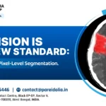
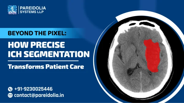
Beyond the Pixel: How Precise ICH Segmentation Transforms Patient Care
In modern healthcare, the fusion of artificial intelligence (AI) and medical imaging is revolutionizing diagnostics and treatment planning. Among the most life-threatening neurological conditions, Intracerebral Hemorrhage (ICH) demands rapid and accurate assessment. The challenge lies in precisely identifying and quantifying hemorrhagic regions within the brain — a process that can make the difference between life and death.
At Pareidolia Systems LLP, we specialize in AI-based ICH segmentation in medical imaging, empowering healthcare professionals with data-driven insights that enhance diagnostic accuracy, streamline workflows, and ultimately improve patient care. Our focus is clear — to go beyond the pixel by turning complex medical images into clinically meaningful data through precise segmentation, annotation, and 3D modeling.
Understanding ICH and Its Clinical Significance
Intracerebral Hemorrhage (ICH) occurs when a blood vessel ruptures within the brain, leading to localized bleeding and pressure buildup. Rapid diagnosis and treatment are crucial to minimize neurological damage and mortality. Traditionally, radiologists rely on CT and MRI scans to identify the affected regions, manually outlining hemorrhages for measurement and evaluation.
However, manual segmentation can be time-consuming, subjective, and prone to variability. In contrast, AI-based ICH segmentation delivers consistency, speed, and high precision — providing clinicians with reliable insights in critical moments.
This technological shift is the foundation of Pareidolia Systems’ mission: transforming diagnostic accuracy through medical image segmentation for stroke care and hemorrhage detection.
Why is accurate ICH Segmentation essential?
ICH segmentation in medical imaging refers to the process of isolating and outlining hemorrhagic areas from CT or MRI scans using algorithmic and manual techniques. It enables:
- Precise measurement of the hemorrhage volume.
- Clear differentiation between healthy and affected brain tissues.
- Quantitative insights for treatment planning and prognosis.
In an era where AI is redefining medicine, deep learning for ICH segmentation enhances the radiologist’s ability to make accurate and timely decisions. Pareidolia Systems provides the critical data backbone required for these models through pixel-perfect annotation and high-quality segmentation.
Pareidolia’s Role in Advancing AI-Based ICH Segmentation
At Pareidolia Systems LLP, we bridge the gap between medical imaging data and actionable clinical intelligence. Our work in AI-based ICH segmentation involves building high-quality, annotated datasets that train and validate AI models for brain hemorrhage detection and analysis.
1. Data Collection and Curation
Our process begins with gathering de-identified and compliant datasets. We ensure that every CT and MRI image is standardized, anonymized, and optimized for use in medical AI development. This foundational step ensures accuracy, privacy, and ethical compliance.
2. Annotation and Labeling Precision
Pareidolia’s expert annotation team marks hemorrhagic regions at the pixel level, ensuring each boundary reflects clinical and anatomical accuracy. This meticulous approach forms the cornerstone for training robust AI algorithms capable of automated brain hemorrhage detection.
3. Advanced Segmentation Techniques
We employ both manual and semi-automated segmentation methods to ensure precision in medical image segmentation for stroke and hemorrhage diagnosis. Our specialists validate each segment through multi-stage quality control, ensuring data consistency across modalities and patient variations.
4. 3D Modeling and Reconstruction
To go beyond traditional 2D interpretation, Pareidolia integrates 3D modeling and visualization of ICH-affected regions. This allows clinicians and researchers to understand the spatial distribution and depth of hemorrhages, supporting better treatment planning and clinical decision-making.
Clinical Impact of ICH Segmentation in Medical Imaging
The clinical impact of ICH segmentation is profound — from faster diagnostics to personalized treatment. Here’s how Pareidolia’s precise segmentation contributes to better outcomes.
1. Accelerated Diagnosis and Triage
AI-assisted ICH segmentation drastically reduces the time required for image evaluation. By automating the detection process, clinicians receive near-instant alerts about potential hemorrhagic areas, allowing faster triage and emergency intervention.
2. Enhanced Diagnostic Accuracy
Manual interpretations can differ between radiologists. Pareidolia’s AI-based segmentation datasets help create models that deliver standardized, reproducible, and objective results, reducing variability in diagnosis.
3. Data-Driven Treatment Planning
With precise volumetric measurements derived from segmentation, doctors can assess the size, shape, and expansion of the hemorrhage with high confidence. These insights guide surgical decisions, medication dosage, and monitoring strategies.
4. Improved Clinical Research
The availability of accurate ICH segmentation data accelerates neuroimaging research. It enables deep learning for ICH segmentation studies, model validation, and cross-institutional collaboration, helping advance new frontiers in stroke and brain injury management.
Pareidolia’s Stepwise Approach to Healthcare AI Development
Pareidolia Systems LLP operates through a structured process that ensures data precision and clinical applicability at every stage.
-
- Stage 1: Data
We draft SOP according to client’s need so that only those clinical data are included which meet the inclusion criteria. All unreliable and inconsistent data are filtered out during this process. - Stage 2: Annotation
Performing detailed medical image annotation, including ICH segmentation and labeling of affected brain structures. - Stage 2A: Segmentation & 3D Modeling
Creating pixel-level segmentations and 3D reconstructions that provide depth and spatial understanding of hemorrhagic regions. - Stage 3: We ensure pixel-perfect annotation and spatially accurate segmentation of intracerebral hemorrhages, enabling AI systems to learn from clinically rich, high-fidelity data.
- Stage 1: Data
This structured approach ensures that Pareidolia’s work remains both technically rigorous and clinically meaningful.
Challenges in AI-Based ICH Segmentation and Pareidolia’s Solutions
1. Data Complexity
Medical imaging data often varies due to equipment differences and patient-specific conditions. Pareidolia addresses this by using standardized preprocessing and multi-level quality control, ensuring uniform segmentation quality.
2. Anatomical Diversity
ICH presentations differ widely in shape, size, and location. Our annotation protocols are designed to adapt to these variations, ensuring that AI models trained on Pareidolia’s data perform reliably across diverse patient profiles.
3. Balancing Automation and Human Expertise
While automation improves speed, expert validation remains essential. Pareidolia blends AI-assisted tools with expert medical annotators, ensuring that segmentation outputs are both efficient and clinically accurate.
4. Ensuring Data Privacy and Security
All medical datasets handled by Pareidolia undergo de-identification and encryption in compliance with healthcare data protection standards. Patient privacy is maintained at every stage of the annotation and segmentation process.
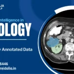
Deep Learning for ICH Segmentation: The Next Frontier
The integration of deep learning in ICH segmentation is redefining neuroimaging analysis. These AI models, trained on Pareidolia’s meticulously segmented datasets, can:
- Detect hemorrhagic regions in real time.
- Predict expansion trends of brain bleeds.
- Assist radiologists in distinguishing between ICH subtypes.
By providing high-quality annotated training data, Pareidolia Systems accelerates the evolution of intelligent imaging tools capable of precision ICH diagnosis using AI.
Why Precision Matters: Beyond the Pixel
Precision in segmentation is more than technical excellence — it’s a clinical necessity. Each pixel accurately identified contributes to a better understanding of the hemorrhage’s extent and impact.
Pareidolia Systems’ approach combines technological innovation with medical understanding, ensuring that every segmentation delivers actionable insight. This philosophy — “We Annotate. You Innovate.” — reflects our mission to empower healthcare AI solutions with meaningful, high-quality data.
The Future of AI in Medical Imaging
The future of medical image segmentation for stroke care lies in automation, integration, and real-time analytics. As AI models become more sophisticated, their ability to assist clinicians in complex neurological diagnoses will only increase.
Pareidolia Systems aims to remain at the forefront of this transformation — bridging the gap between raw imaging data and actionable medical intelligence. Through AI-based ICH segmentation, 3D modeling, and data-driven research support, we continue to redefine how imaging data contributes to clinical excellence.
In healthcare, every second and every pixel counts. Precise ICH segmentation in medical imaging is transforming how clinicians diagnose, plan, and treat patients suffering from intracerebral hemorrhage.
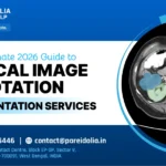
By combining expertise in AI-based data annotation, medical image segmentation, and 3D modeling, Pareidolia Systems LLP enables medical institutions, researchers, and AI developers to unlock new dimensions of diagnostic precision and patient care.
At Pareidolia, our vision is simple yet powerful:
We Annotate. You Innovate.
Through accuracy, consistency, and clinical understanding, we move beyond the pixel — transforming raw data into lifesaving insight.