 Feb 14 2025
 AI-Powered Health Data Collection
What is deep learning in radiology?
In today’s fast-paced world, staying updated with AI technology—especially deep learning in radiology—is no longer optional. Whether you’re aiming to advance your career in the field or simply looking to expand your knowledge, understanding these advancements is essential. Missing out on this technology means falling behind in an industry that’s rapidly evolving and shaping the future of medical imaging.
Deep Learning in Radiology is a leader in this battle with technology that was able to make fast advancements and revolutionize healthcare. With this advanced technology, AI transforms how radiological data is analyzed & interpreted.
But how exactly is radiology, and how does deep learning enhance its potential in healthcare? After finishing this article, you will get answers to all your queries.
What Is Radiology?
Radiology is an imaging technique to diagnose and treat diseases within the body. Some methods involve X-rays, MRI (Magnetic Resonance Imaging), CT (Computed Tomography) scans, ultrasound, and PET (Positron Emission Tomography) scans. Radiologists read these images to identify abnormalities and help doctors decide on treatment options.
In radiology, Mostly There are two broad categories:
- Diagnostic Radiology: Center for Diagnosing Disease Imaging Disease through CT scans, X-rays, and MRIs.
- Interventional Radiology: IOf minimally invasive procedures, such as catheters, guided by imaging techniques or performing biopsies.
Why Is Radiology Important in Healthcare?
Radiology plays a very important role in modern healthcare due to several reasons:
- Early Diagnosis: Radiology helps in early disease detection, and imaging techniques lay the groundwork for patients to improve their outcomes and survival rates.
- Treatment Monitoring: Radiology enables doctors to track the progress of treatments and change them if necessary. For instance, to evaluate tumor regression during chemotherapy.
- Non-Invasive Procedures: Imaging is typically a non-invasive procedure, making it less risky and more comfortable for patients than surgical approaches.
- Precision Medicine: By processing imaging data, radiological images identify detailed information about a specific structure and its composition, aiding in the design of personalized therapy for the patients.
- Surgical Planning: By processing imaging data, radiological images identify detailed information about a specific structure and its composition, aiding in the design of personalized therapy for the patients.
An Introduction to Deep Learning
So, deep learning is a subset of artificial intelligence in which neural networks are trained on large amounts of data to identify patterns and make predictions. Deep learning recently made its way to the field of radiology, effecting a change that presented medical imaging as an efficient, accurate, and automated process. Over time, deep learning in radiology makes the process more easy & efficient. Let’s get an idea of the domains where the power of deep learning is unprecedentedly being used.
Image Classification
Image Classification is The Process Where Deep learning algorithms can find patterns in radiological images of the human body to detect normal and abnormal findings. For instance, AI Techniques in Radiology can recognize different types of chest X-rays to see which have pneumonia or CT scans to spot cancerous lesions.
These classifications are made based on vast training performed on labeled datasets, allowing the models to learn and identify even minor changes.
Object Detection
Object Detection is another major contribution. It helps radiologists concentrate on the areas of interest (such as lesions, fractures, and tumors) by using deep learning models to find the areas of interest. This ability can be of great help to uncover minute or more easily missed anomalies, enhancing diagnostic accuracy. Artificial Intelligence in Medical Imaging is changing the way we Detect any radiological Object.
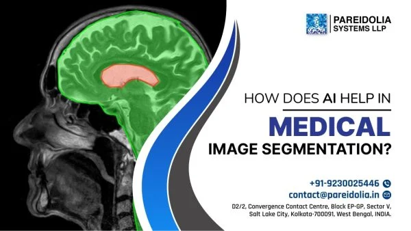
Semantic Segmentation
Semantic segmentation is a process of labeling each pixel of an image with its category. For example, identifying tissues, organs, and pathological regions within a CT image. It Gives a more detailed analysis, which is critical in tasks like tumor boundary delineation or organ volume estimation.
Instance Segmentation
Aspectimg/ Compared to semantic segmentation, in image instance segmentation, a specific object can be retrieved individually from an image. This is vital for applications like measuring tumor volume, counting lesions, or detecting branching structures in tissues with overlapping anatomies.
Data
AI Applications in Radiology require data to train a relatively complex network. However, this data in question is, in the case of radiology, derived from medical images and patient records as well as expert annotations. However, there are challenges and opportunities in working with this data.
Convolutional Neural Networks
Deep learning in radiology is powered by Convolutional Neural Networks (CNNs). Convolutional Neural Networks (CNNs) are particularly well-suited for image data, as they use multiple layers of neurons to extract increasingly complex hierarchical features from images, from edges and textures to complex patterns. CNN played a key role in opening up a range of applications from image classification to segmentation with algorithms that won many awards in state-of-the-art medical imaging and beyond. The Top features of CNN are:
- Effectively use convolutional layers to extract features from image data
- Versatile – Can be used for all types of tasks – segmentation, detection, classification
Toward Deeper Networks
The use of Deep Learning for Medical Image Analysis has become one of the most efficient strategies, which consists of several layers of neurons, increasing the potential for models to learn complex features in medical images.
These networks can help in dealing with complex datasets in radiology use cases. You will often come across examples like ResNet and DenseNet architectures that can revolutionize the approach to various challenges like vanishing gradients or overfitting and hence make a deep network more applicable in medical settings. How It Helps Us:
- Solve complicated problems using several levels of hierarchy.
- However, as ResNet also had skip connections, there were innovations to help with vanishing gradients as well.
Deep Learning in Radiology vs. “Traditional” Machine Learning in Radiology
- Traditional Machine Learning: Dependent on handcrafted feature selection, which necessitates domain knowledge to manually engineer features. These functions tend to perform poorly on high-dimensional data, such as medical images.
- Deep Learning: Enables even unseen data to train, enabling powerful models. Also, automation lowers the time taken than the human factor.
- Complex Data: Deep learning, with its multilayered network,s can easily handle unstructured and high-dimensional datasets like 3D medical imaging better than the conventional methods.
- Scalability: While traditional machine learning needs to have features redesigned as data increases in size, deep learning can just be scaled up on more data.
State-of-the-Art Classification: Deep Learning in Radiology
Deep learning models are excellent at image classification, assisting with diagnostic diseases such as pneumonia, cancer, or cardiovascular anomalies. By being trained to tell the difference between normal and pathological findings, these models have reduced diagnostic error rates and improved throughput.
AI techniques for classifying mammograms to highlight early-stage breast cancer with high sensitivity can help radiologists make timely judgments about a patient’s condition.
Segmentation
AI-Power Image segmentation using artificial intelligence automates the process of delineating structures in medical images. It is specifically beneficial for applications such as tumor segmentation, organ boundary mapping, and blood vessel tracking. Automated Medical Image Segmentation is an immense time saver and leads to better consistency and accuracy compared to manual approaches.
Detection
In AI in Diagnostic Imaging, detection models examine images for abnormalities such as microcalcifications in mammograms, nodules in chest X-rays, or fractures in bone scans. The best of these models can often detect subtle or rare findings better than humans and so promise to be particularly useful in busy clinical settings.
The Benefits of Using Deep Learning in Radiology
- Improved Accuracy: It helps reduce errors and increases diagnostic confidence by identifying likely abnormalities that might be omitted by a human eye.
- Time Efficiency: Reduces manual effort, allowing radiologists to concentrate on complex cases and patient-oriented work.
- Cost-Effectiveness: Decreases the burden on healthcare costs through optimized workflows and eliminating unnecessary testing.
- Enhanced Accessibility: Provides AI-powered diagnostic tools that bridge the gap in underserved areas where radiologists are unavailable
- Scalability: That can handle high volumes of data without affecting quality, perfect for large patient-load institutions
- Personalized Insights: Offers more granular and patient-specific information that can assist with precision medicine.
Other Tasks in Radiology
Image Registration:
Image registration (aligning images from a few modalities like MRI, PET, etc.) This process adds another layer of information to the insight into the patient’s ailments to make a deeper look into the condition to make a better diagnosis and a thorough plan for upcoming therapy.
Image Generation/Reconstruction:
Some image generation/reconstruction algorithms are developed to upscale images (for example, making a low-resolution scan cleaner and more accurate) or to fill in missing portions of an image. This is especially useful in minimizing radiation exposure by allowing excellent-quality imaging with lower doses.
Image Enhancement:
Image enhancement allows for the better visualization of certain features, such as blood vessels or soft tissue contrast accumulation. These techniques help radiologists identify abnormalities and make accurate diagnoses faster.
Main Challenges and Pitfalls in the Development of Deep Learning Algorithms
Despite its promise, the integration of AI in Medical Imaging faces several challenges:
- Data Privacy: Thus, members will get their sensitive patient information protected by model training encryption and comply with regulations like HIPAA.
- Data Quality: Data must be accurate, diverse, and free from bias to prevent skewed results and ensure fairness.
- Interpretability: One major hurdle remains how to make AI choices interpretable to clinical staff since many deep learning models are dubbed “black boxes.”
- Regulation and Approval: A lengthy and expensive process is often needed to satisfy the stringent regulatory requirements to be approved for clinical use.
- Integration: It takes a great deal of training, infrastructure, and adaptation to seamlessly integrate AI tools into existing workflows.
Future of Deep Learning in Radiology
Looking ahead, the future of Deep Learning in Radiology is bright:
- Personalized Medicine: The AI-imbued insights will allow for treatments designed and targeted to the specific patient on a personalized basis.
- Predictive Analytics: Predictive AI will also enable proactive healthcare by predicting. How individuals will respond to treatments and how diseases progress.
- Remote Diagnostics: By integrating AI into telemedicine, you can reach remote and hinterland areas with quality healthcare.
- Collaborative AI Systems: Supporting the work radiologists do with complementary tools of AI will promote a more collaborative approach to diagnosis and treatment.
- Continuous Learning: As new data is established, which seems to be occurring at a rapid pace, models that are trained on it will naturally do better. Becoming more attuned to the current dynamics of the data and tools.
- AI-Powered Image Segmentation: Ongoing developments will enhance segmentation methods, enabling greater precision and utility in diverse implementations.
Deep Learning in Radiology is more than a technological revolution; it is a paradigm shift in healthcare delivery. With the implementation of AI-enabled solutions, the field of radiology is achieving greater accuracy, accessibility, and efficiency. If you use artificial intelligence wisely. It will not only be a new challenge but also a challenge that will lead to innovations and, in the future. Change the health condition forecasting method.
By The Use of Machine Learning in Medical Imaging just made a revalorization in the Medical World. Also, There is still a lot of Improvement & Growth. But with the right intent, we get the massive success we are looking for. Join us & Get Our Advanced AI-Power Medical Service in Healthcare.