 Jan 13 2026
 AI in Neurological Imaging
Artificial intelligence in neurology is transforming how clinicians detect, diagnose, and monitor neurological disorders
AI in neurology is transforming how clinicians detect, diagnose, and monitor neurological disorders. From early signs of Alzheimer’s to subtle markers of multiple sclerosis, AI-driven models promise faster and more accurate insights than traditional imaging workflows.
However, the success of neurology AI does not begin with algorithms—it begins with high-quality medical image annotation.
One of the biggest bottlenecks in building reliable neurological AI systems is low-contrast MRI segmentation. Early-stage neurological abnormalities often appear faint, ambiguous, and difficult even for experienced radiologists to delineate. Teaching AI to “see” what is barely visible requires precise annotation, expert-guided segmentation, and rigorous quality control.
At Pareidolia, we specialize in annotating the unseen—helping AI systems learn from complex, low-signal medical images through advanced medical image segmentation, expert annotation workflows, and clinical-grade quality assurance.
Why Low-Contrast MRI Is a Critical Challenge in Neurology AI?
Magnetic Resonance Imaging (MRI) is the gold standard for neurological imaging, but not all MRIs are created equal. In early-stage neurology, disease markers often present as:
- Minimal intensity differences between tissues.
- Subtle lesions with unclear boundaries.
- Early neurodegenerative changes that blend into normal anatomy.
These challenges make MRI image segmentation significantly harder compared to high-contrast modalities.
The Problem with Early-Stage Neurological MRI Data behind AI models
Early-stage neurological MRI segmentation faces several obstacles:
- Poor gray-white matter contrast.
- Low signal-to-noise ratio (SNR).
- Diffuse or non-localized abnormalities.
- High inter-patient variability.
- Scanner and protocol inconsistencies.
For AI models, these challenges translate into misclassification, poor generalization, and unreliable predictions—unless the training data is annotated with exceptional precision.
This is where medical image annotation for neurology AI becomes mission-critical.
Why Annotation Quality Determines AI Performance in Neurology?
AI models are only as good as the data they learn from. In neurological imaging, annotation errors—however small—can have major downstream consequences.
Annotation Is Not Just Labeling
Effective annotation of medical images involves:
- Precise boundary delineation.
- Anatomical consistency across slices.
- Pathology-aware labeling.
- Cross-validation by domain experts.
For low-contrast MRI, this process requires human-in-the-loop expertise, advanced segmentation strategies, and robust quality control systems.
At Pareidolia, annotation is treated as a clinical-grade process, not a mechanical task.
Medical Image Segmentation: The Foundation of Neurology AI
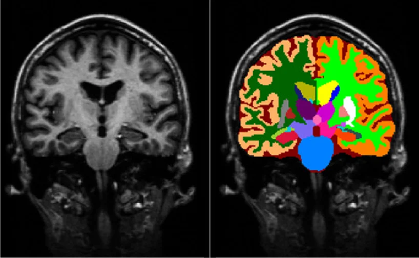
Medical image segmentation is the process of partitioning MRI scans into meaningful regions—such as brain structures, lesions, or tissue classes.
Why Segmentation Matters in Neurology AI
Accurate segmentation enables:
- Early disease detection using MRI.
- Quantitative analysis of brain structures.
- Progression tracking in longitudinal studies.
- Training of robust neurology AI models.
Without reliable segmentation, even the most advanced AI architectures fail to deliver clinical value.
Low-Contrast MRI Segmentation Requires Specialized Expertise
Generic segmentation pipelines often fail in early neurology use cases. Low-contrast MRI segmentation demands:
- Expert-guided annotation strategies
- Multi-planar and multi-sequence analysis.
- Context-aware boundary decisions.
- Iterative validation loops.
Pareidolia’s segmentation workflows are designed specifically to handle low-visibility neurological features, ensuring AI-ready datasets.
Annotation of Medical Images for Neurology AI
Annotation is the bridge between raw MRI scans and intelligent AI systems.
What Makes Neurology Annotation Unique?
Neurological image annotation differs from other domains due to:
- Complex brain anatomy.
- Subtle pathological changes.
- High clinical risk of mislabeling.
- Need for anatomical and pathological coherence.
Our medical image annotation for AI workflows combine:
- Trained medical annotators.
- Radiology-guided protocols.
- Neurology-specific annotation standards.
This ensures datasets are suitable for neurology AI training data pipelines.
Brain MRI Segmentation Techniques Used in Early-Stage Neurology
Effective brain MRI segmentation techniques for low-contrast data rely on a hybrid approach.
Manual and Semi-Automated Segmentation
While automated tools accelerate workflows, early-stage neurological MRI still benefits from:
- Manual fine-tuning by experts.
- Semi-automated segmentation with human validation.
- Slice-by-slice anatomical consistency checks.
At Pareidolia, this hybrid approach is powered by both clinical expertise and platform fluency:
- Deep familiarity with normal and pathological brain anatomy across neurological conditions
- Hands-on experience with leading segmentation platforms, including Redbrick, Mimics, ITK-SNAP, and 3D Slicer
- Ability to adapt workflows across tools while maintaining anatomical and labeling consistency
- Expert oversight that ensures every pixel reflects true clinical meaning, not just algorithmic output
MRI Preprocessing for AI Models: Preparing Data for Learning
Before annotation begins, MRI data must be prepared so that subtle neurological structures are clearly visible and consistent across scans. In low-contrast brain MRI, poor preprocessing can cause noise or scanner artifacts to be mislabeled as anatomy.
Key MRI Preprocessing Steps
Effective MRI preprocessing for AI models includes:
- Noise reduction.
- Intensity normalization.
- Bias field correction.
- Image registration and alignment.
These steps ensure that annotators label true clinical features, not imaging artifacts—resulting in more reliable AI training data.

3D Model Creation: Adding Depth to Neurology AI
Beyond 2D segmentation, 3D model creation plays a vital role in neurological AI applications.
Why 3D Models Matter?
3D reconstructions enable:
- Better spatial understanding of brain structures.
- Volumetric analysis of lesions.
- Improved AI interpretability.
- Enhanced visualization for clinical and research use.
Pareidolia converts segmented MRI data into accurate 3D anatomical models, empowering advanced neurology AI systems.
Quality Control in Medical Imaging: Ensuring Clinical-Grade Data
In neurology AI, annotation errors can lead to false diagnoses or missed early warnings. That’s why quality control in medical imaging is non-negotiable.
Pareidolia’s Multi-Layer Quality Assurance
Our QC framework includes:
- Multi-review annotation validation.
- Inter-annotator consistency checks.
- Radiology and neurology expert audits.
- Statistical accuracy benchmarking.
This guarantees accurately annotated neurology imaging datasets that adhere to research and clinical requirements
AI in Neurological Imaging: From Research to Real-World Impact
High-quality annotated data enables AI systems to:
- Detect neurological disorders earlier.
- Support clinical decision-making.
- Reduce radiologist workload.
- Improve patient outcomes.
With accurate neurological disorder detection AI, subtle early-stage signals can be identified before symptoms escalate.
Why Pareidolia Is Trusted for Neurology AI Annotation?
Pareidolia is more than an annotation provider—we are a medical imaging partner.
What Sets Pareidolia Apart
- Deep expertise in neurology and radiology
- Specialized workflows for low-contrast MRI segmentation
- End-to-end services: segmentation, annotation, 3D modeling, QC
- AI-ready datasets designed for scalability and accuracy
We understand that artificial intelligence in neurology depends on precision, not assumptions.
The Future of Early-Stage Neurology AI Starts with Better Annotation
As AI continues to redefine neurological care, the demand for high-quality, expertly annotated MRI datasets will only grow.
Early-stage neurology AI cannot rely on visible abnormalities alone—it must learn from the unseen, the subtle, and the ambiguous.
At Pareidolia, we help AI models see what is easy to miss.
By combining medical image segmentation, annotation of medical images, 3D model creation, and quality control in medical imaging, we enable neurology AI to move from potential to practice.
Ready to Build Better Neurology AI?
If your AI models rely on MRI data and early-stage neurological insights, Pareidolia provides the expertise, precision, and scalability you need.
Annotate smarter. Segment deeper. Detect earlier.