
 Mar 20 2025
 AI-Powered Health Data Collection
Achieving Accurate Diagnosis of Cerebral Stenosis: The Power of Segmentation & Annotation in CT Imaging
Cerebral stenosis is essentially the narrowing of blood vessels in the brain, which can result in stroke and a decline in cognitive function. While early detection is vital, new developments in CT imaging have enabled identification. However accurate analysis requires specialized experience.
This blog highlights the importance of segmentation and annotation services in analyzing CT images for cerebral stenosis. Our expert solutions offer cost-effective, high-accuracy results, aiding both early detection and AI-ML research to enhance healthcare outcomes.
What is Cerebral Stenosis?
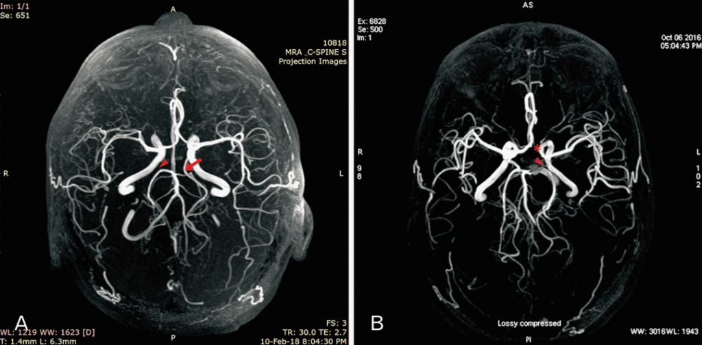
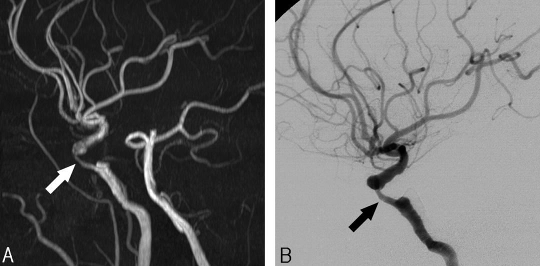
Cerebral stenosis is the narrowing of arteries that supply oxygen-rich blood to the brain, often due to plaque buildup. This condition can develop silently, with minimal symptoms, making early detection through CT imaging essential for preventing severe complications.
Why is CT Imaging Essential for Stenosis Detection?
CT scans provide high-resolution images of the brain, making it easier to detect blood vessel narrowing. However, due to the complexity of brain structures, advanced segmentation and annotation techniques are needed for precise diagnosis.
By isolating key areas, segmentation and annotation simplify the analysis, ensuring accurate detection of stenosis. These techniques improve diagnostic accuracy, enabling healthcare professionals to make informed treatment decisions.
Segmentation & Annotation Services: Enhancing Stenosis Detection
1. Segmentation:
Segmentation divides CT images into regions of interest, isolating blood vessels and detecting narrowed sections caused by plaque buildup.
Our advanced image processing solutions ensure precise segmentation, offering:
- High-resolution analysis for accurate identification of affected vessels.
- 3D segmentation services providing a detailed view of blood flow restrictions.
- Enhanced early detection of stenosis for proactive healthcare solutions.
At Pareidolia Systems LLP, we empower healthcare professionals with cutting-edge imaging services for better diagnostics and patient care. This process enables early diagnosis and better treatment outcomes for patients.
2. Annotation:
Annotation involves labeling key features in segmented images, such as the location, size, and severity of stenosis. Accurate annotation benefits both medical professionals and AI systems, aiding in automatic stenosis detection.
Benefits of precise annotation:
- Improved insights for healthcare professionals.
- Enhanced AI model training for automatic stenosis detection.
- More accurate recognition of stenosis patterns in future scans.
Empowering AI-ML Model Development for Stenosis Detection
With AI-ML integration in healthcare, early detection and diagnosis of cerebral stenosis are becoming more efficient. AI models trained on high-quality annotated CT images can identify stenosis faster, reducing misdiagnosis risks.
Our segmentation and annotation services support AI-ML research by providing labeled datasets essential for training advanced models. We collaborate with leading companies to develop AI systems that:
- Automatically detect stenosis in CT scans
- Predict severity based on vessel narrowing
- Identify early signs of plaque buildup
With high-accuracy data, AI models continuously improve, leading to faster diagnoses, precise results, and enhanced patient care.
Why Choose Our Expertise?
1. High-Accuracy Detection:
We specialize in precision-driven segmentation and annotation, ensuring reliable CT image analysis. Our accuracy enhances both medical diagnosis and AI model training.
2. Cost-Effective Solutions:
We provide high-quality annotated datasets at affordable prices, making AI-ML model development accessible to healthcare and research organizations.
3. Experienced Medical Imaging Specialists:
Our team comprises experts in segmentation algorithms, deep learning, and medical image analysis, ensuring precise CT image processing.
4. Fast Turnaround Time:
We deliver quick and efficient segmentation and annotation services, accelerating AI research and diagnostic workflows.
Proven Success with Industry Leaders
We collaborate with top healthcare companies to advance AI-driven detection of cerebral stenosis. Our accurate annotated datasets help develop AI models that:
- Improve diagnostic workflows.
- Enable early intervention.
- Enhance healthcare efficiency.
Shaping the Future of Cerebral Stenosis Detection with AI
Early and accurate detection of cerebral stenosis is vital for preventing severe neurological consequences. Our segmentation and annotation services empower healthcare providers, researchers, and AI developers to enhance stenosis detection capabilities.
By partnering with us, you gain access to expert-driven, cost-effective solutions that support AI-ML advancements in healthcare. Together, we can create a future where early detection leads to optimized treatment and improved patient outcomes.
What is Segmentation & Annotation in CT Imaging?
Segmentation in medical imaging refers to the process of isolating and highlighting specific areas of a scan, such as blood vessels or abnormal tissues. Annotation involves labeling these segmented areas to help radiologists and AI-powered systems quickly identify problem zones.
By applying AI-driven segmentation and annotation, CT imaging becomes far more efficient in detecting cerebral stenosis, ensuring faster diagnoses and better treatment outcomes.
How Segmentation & Annotation Improve Diagnosis
1. Clearer, More Accurate Imaging
Standard CT scans can be difficult to interpret, especially when dealing with intricate brain structures. AI-based segmentation enhances image clarity, allowing doctors to easily distinguish between normal and narrowed blood vessels.
2. Faster & More Precise Diagnosis
Manual review of CT scans takes time and is prone to human error. Automated segmentation tools quickly highlight areas of concern, helping radiologists diagnose cerebral stenosis with greater accuracy and speed.
3. Quantifying Vessel Narrowing for Better Treatment Planning
Segmentation helps measure the exact degree of narrowing in blood vessels, allowing doctors to determine whether a patient needs:
- Medication to manage symptoms.
- Minimally invasive procedures like stenting.
- Surgery for severe cases.
4. Enhancing Workflow for Radiologists
AI-powered annotation reduces the time radiologists spend analyzing images, enabling them to focus on treatment planning and patient care. This leads to faster results and improved efficiency in hospitals and clinics.
AI & Machine Learning: The Future of CT Imaging
With artificial intelligence (AI) and machine learning (ML), CT imaging is becoming more powerful than ever. These technologies can:
- Detect even the smallest abnormalities in blood vessels.
- Provide real-time analysis for faster decision-making.
- Continuously improve accuracy through data-driven learning.
AI is transforming how we diagnose cerebral stenosis, making early detection easier and reducing the risks of stroke and brain damage.
The Future of Cerebral Stenosis Diagnosis
As CT imaging technology advances, segmentation and annotation tools will continue to evolve. Future innovations may include:
🔹 Real-time 3D visualization of brain arteries.
🔹 Predictive analytics to assess stroke risks.
🔹 AI-assisted decision-making for customized treatment plans.
CT imaging remains the gold standard for diagnosing cerebral stenosis, but AI-powered segmentation and annotation are taking it to the next level. These technologies ensure faster, more accurate, and more efficient diagnoses, helping doctors make informed treatment decisions and ultimately saving lives.
💡 Stay ahead of the curve with AI-driven medical imaging and improve patient outcomes today!
Contact us today to discover how our segmentation and annotation services can enhance your research, AI models, and healthcare innovations.
Latest Posts
-
 Artificial intelligence in neurology is transforming how clinicians detect, diagnose, and monitor neurological disorders
 -
 The Blueprint for Better Stroke Care:How Pareidolia’s Precise Annotations Are Training Life-Saving AI
 -
 3D Printing in Neurosurgery: How Neurologists Use 3D Models for Aneurysm Surgery Planning