 May 22 2025
 medical image analysis
What are the Latest Advancements in Medical Imaging Analysis?
Medical image analysis has witnessed remarkable progress in recent years, driven primarily by advancements in artificial intelligence (AI) and machine learning (ML). These technologies have revolutionized how researchers and clinicians interpret complex medical data, enhancing diagnostic accuracy and enabling personalized treatment strategies. In this blog, we explore the latest advancements in medical imaging analysis, focusing on core areas such as medical image segmentation, annotation, 3D model creation, and quality control — all critical components in AI-powered healthcare solutions.
Introduction to Medical Imaging Analysis
Medical imaging analysis involves processing and interpreting images generated using equipment such as MRI, CT scan, X-ray, and ultrasound. Traditionally, this was a manual, time-intensive process prone to variability and human error. However, AI-driven methods are transforming this domain by automating image interpretation with precision and speed.
A. Medical Image Segmentation: The Cornerstone of AI Analysis
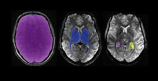
Medical image segmentation refers to the process of partitioning an image into meaningful regions, such as identifying organs, tumors, or lesions. This step is crucial because it enables AI models to focus on relevant structures, improving disease detection and treatment planning.
Latest Advances:
- Deep Learning Architectures: Models like U-Net, Mask R-CNN, and Transformers like Sega Former have become standard for segmentation tasks, offering unprecedented accuracy on complex datasets. These tools can perform semantic as well as instance segmentation.
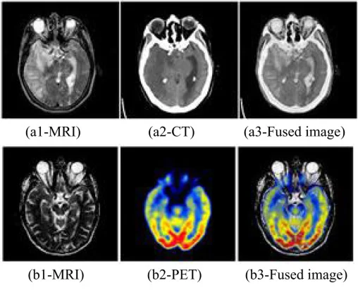
- Multi-modal Segmentation: Combining data from multiple imaging modalities (e.g., PET, MR & PET CT) improves segmentation robustness, enabling more comprehensive analysis.
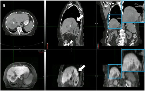
- 3D Segmentation: Advances in volumetric segmentation allow entire organs or tumors to be segmented in three dimensions rather than slice-by-slice, enhancing the anatomical understanding.
B. Annotation of Medical Images: Building Reliable AI Datasets
High-quality annotated datasets are the backbone of any supervised AI system. Annotation involves labeling regions or features in medical images, which trains AI algorithms to recognize and interpret similar patterns automatically. Building reliable AI datasets is essential for AI models to accurately detect patterns, improve diagnoses, personalize treatments, and reduce errors, ultimately enhancing patient outcomes and safety. Additionally, comprehensive datasets help ensure fairness by representing diverse populations, support regulatory compliance, and drive innovation and trust in AI-driven healthcare solutions.
Latest Advances:
- Semi-Automated Annotation Tools: New software solutions assist experts by providing initial labels that can be quickly refined, drastically reducing annotation time while maintaining accuracy.
- Crowdsourcing & Expert Review Integration: Combining crowd-labeled data with expert validation optimizes both scale and quality of datasets.
- Standardization Efforts: Consistent annotation protocols improve dataset interoperability, facilitating better model generalization across institutions.
C. 3D Model Creation: From Flat Images to Realistic Anatomy
Creating 3D models from medical images adds a spatial dimension critical for diagnostics, surgical planning, and simulation.
Latest Advances:
- AI-Assisted 3D Reconstruction: Deep learning models now automate complex 3D reconstruction from 2D slices, making realistic anatomical models accessible faster than ever.
- Interactive Visualization Tools: Augmented reality (AR) and virtual reality (VR) platforms integrate 3D medical models for immersive clinical review and training.
- Patient-Specific Modeling: Personalized 3D models improve pre-operative planning and customized implant design.
- 3D Printing Applications: These digital models can also be used for 3D printing, enabling surgeons to rehearse complex procedures and helping patients better understand their treatment.
D. Quality Control in Medical Imaging: Ensuring Trustworthy AI Outcomes
Quality control (QC) is often overlooked but is essential for reliable medical image analysis. QC ensures the integrity, consistency, and accuracy of image data and annotations before they used in AI model training or clinical applications.
Latest Advances:
- Automated QC Pipelines: AI-powered systems detect and flag low-quality images or annotation errors, maintaining dataset standards.
- Hybrid QC Approaches: Combining automated tools with human expert review achieves optimal balance between efficiency and accuracy.
- Real-Time QC in Clinical Settings: Emerging solutions integrate QC checks during imaging acquisition, reducing the need for repeat scans.
E. Software for Medical Image Analysis: Tools Shaping the Future
The software landscape for medical image analysis is evolving rapidly, incorporating AI algorithms, cloud computing, and user-friendly interfaces.
Latest Advances:
- Open-Source Platforms: Tools like 3D Slicer and ITK-SNAP empower researchers to perform segmentation and annotation with customizable pipelines.
- Commercial AI Suites: Companies like Siemens Healthiness and GE Healthcare offer integrated AI-powered analysis suites tailored for clinical workflows.
- Cloud-Based Annotation and QC: Cloud platforms enable collaborative annotation and real-time quality control, speeding up dataset preparation and supporting federated learning and multi-institutional research collaborations.
Medical imaging analysis is at the forefront of AI-driven healthcare innovation. Advances in segmentation, annotation, 3D modeling, and quality control are collectively enabling researchers and clinicians to harness medical images like never before. For AI & ML researchers, understanding and integrating these advancements is crucial to developing robust, clinically viable models that can improve patient outcomes worldwide.
At Pareidolia Systems LLP, we specialize in delivering end-to-end AI solutions encompassing all these critical aspects and help you accelerate development, reduce deployment friction, and meet clinical demands with confidence— Whether you’re building diagnostic tools, surgical planning platforms, or training data pipelines, our end-to-end expertise helping you bridge the gap between raw medical images and actionable AI insights.